Method of evaluating immunosuppression

a immunosuppression and immunosuppression technology, applied in the field of immunosuppression determination, can solve the problems of increasing the rate of infections and cancers, increasing the risk factors for cardiovascular risk factors and bone diseases, and life-long anti-rejection therapy has many adverse effects, so as to achieve less nuclear translocation of nfb, small similarity score, and high negative similarity score

Inactive Publication Date: 2013-07-18
DAVIDOVICH DONNA
View PDF1 Cites 3 Cited by
  • Summary
  • Abstract
  • Description
  • Claims
  • Application Information

AI Technical Summary

Benefits of technology

[0044]In one embodiment, after contacting the cells with the activating agent, cells are immunophenotyped with commercially available fluorescently labeled antibodies, after which the red blood cells are removed from the whole blood sample. Red blood cells can be removed using conventional techniques, such as by lysing using a hyptonic solution under conventional conditions which does not also result in lysis of the activated cells. In one embodiment, a commercially available lyse / fix solution (Becton Dickenson) can be used.
[0045]In one embodiment, prior to determining nuclear NFκB, the cells in which the nuclear NFKB is to be determined are incubated with an antibody specific to a relevant NFKB subunit, wherein the antibody is conjugated to a fluorescent marker. In a similar embodiment, prior to determining nuclear NFAT, the cells in which nuclear NFAT is to be determined are incubated with an antibody specific to a relevant NFAT, wherein the antibody is conjugated to a fluorescent marker. In various embodiments, nuclear NFκB and NFAT can be determined concurrently, or sequentially, and for the same cell population, or for separate cell populations. For certain applications, the antibody directed to a NFκB subunit can be conjugated to with a different fluorescent marker than the antibody directed to NFAT so that they can be separately identified and measured.
[0046]In more detail, in various embodiments of the invention, the relative amounts of nuclear NFκB and / or nuclear NFAT can be determined in activated and / or non-activated samples by analysis with commercially available devices and / or systems that can differentiate and quantify the nuclear and cytoplasmic NFκB and / or NFAT, such as by a variety of digital microcopy-based imaging techniques. For example, activated and / or non-activated preparations of cells could be fixed and analyzed using detectably labeled antibodies to NFκB (such as to p65) and / or NFAT, as applicable, and known reagents to stain or otherwise identify the nucleus such that the nuclear (and if desired cytoplasmic) NFκB, and / or NFAT, as applicable, can be distinguished from one another. Suitable nuclear stains include but are not limited to 4′,6-diamidino-2-phenylindole (DAPI), Hoechst stains, Haematoxylin, Safranin, Carmine alum, and DRAQ5.
[0047]In one embodiment, nuclear NFKB and / or nuclear NFAT can be determined using imaging flow cytometry. For example, the amount of nuclear NFKB in activated and non-activated cells can be determined for CD3+, CD4+, CD8+, and / or CD20+ cells using detectably labeled antibodies directed to the CD3+, CD4+, CD8+ and / or CD20+ molecules, as well as detectably labeled antibodies to NFκB. Similarly, the amount of nuclear NFAT in activated and non-activated cells can be determined for CD3+, CD4+, CD8+, and / or CD20+ cells using detectably labeled antibodies directed to the CD3+, CD4+, CD8+ and / or CD20+ molecules, as well as detectably labeled antibodies to NFAT. The nuclei of the cells can be simultaneously visualized using a suitable nuclear stain that can be detected by an imaging flow cytometer. In one embodiment, the nuclear stain is DAPI. In another embodiment, the nuclear stain is DRAQ5.
[0048]In certain aspects of the invention, determining nuclear NFκB and / or nuclear NFAT is performed using an imaging flow cytometer. In one embodiment, the imaging flow cytometer is an imaging flow cytometer device described in U.S. Pat. No. 7,522,758, the disclosure of which is hereby incorporated by reference. Such image flow cytometers are able to quantitatively measure the relative proportion of NFκB and / or NFAT that is located in the cytoplasm versus the amount in the nucleus. This proportion allows for measuring, among other parameters, the relative activation state of a given cell population between two samples obtained from two different patients or the same patient at different times obtained under differing clinical circumstances. For example, by dual staining for p65 or NFAT1 and cell surface markers such as CD4, CD8, CD19 / 20 and CD16, the image flow cytometer can measure the relative NFkB and NFAT activation in T helper cells, T cytotoxic cells, B cells and Natural Killer cells, respectively. The ability to assess activation states of multiple different cell types represents a major advance compared to the CD4 limited ImmuKnow assay. Thus, in one embodiment, the invention provides for determining a composite result obtained from all or a subset of cell types. The resting state compared to the activated state of peripheral blood lymphocytes (PBL) may accurately reflect in vivo events such as infection and rejection.
[0049]In one embodiment, a relative amount of NFκB and / or NFAT present in the nucleus can be represented by a similarity score determined using an imaging flow cytometer system such as that described in U.S. Pat. No. 7,522,758. In general, the smaller the similarity score, the less nuclear translocation of NFκB and / or NFAT, and vice versa. More specifically, the similarity score is considered to be a log transformed Pearson's Correlation coefficient of the pixel by pixel intensity correlation between the NFκB and / or NFAT and nuclear stained (i.e., DRAQ5 image) which is calculated as a quantifiable parameter for the degree of NFκB and / or NFAT translocation to the nucleus. The similarity score (+ or −) is determined from the slope of the regression line while it takes its value from how well the individual pixel data points fit the regression line (Pearson correlation). A very low degree of nuclear translocation yields a highly negative similarity score while a very high degree of nuclear translocation yields a highly positive similarity score. It will therefore be recognized that, in one embodiment a low degree of nuclear translocation can have anti-similar p65 and DRAQ5 images, while similar p65 and DRAQ5 images can yield a positive similarity score. Thus, in one embodiment, following stimulation, a negative similarity score obtained using an imaging flow cytometer system is indicative of excessive immune suppression, while a highly positive similarity score obtained using an imaging flow cytometer system is indicative of insufficient immune suppression. A standardized similarity score or ranges of similarity scores can accordingly be used as a control when performing the method of the invention.

Problems solved by technology

Potentially life-long anti-rejection therapy has many adverse consequences including increased rates of infections and cancers, worsening cardiovascular risk factors and bone disease.

Method used


Image

  • Method of evaluating immunosuppression
  • Method of evaluating immunosuppression
  • Method of evaluating immunosuppression

Examples


example 1

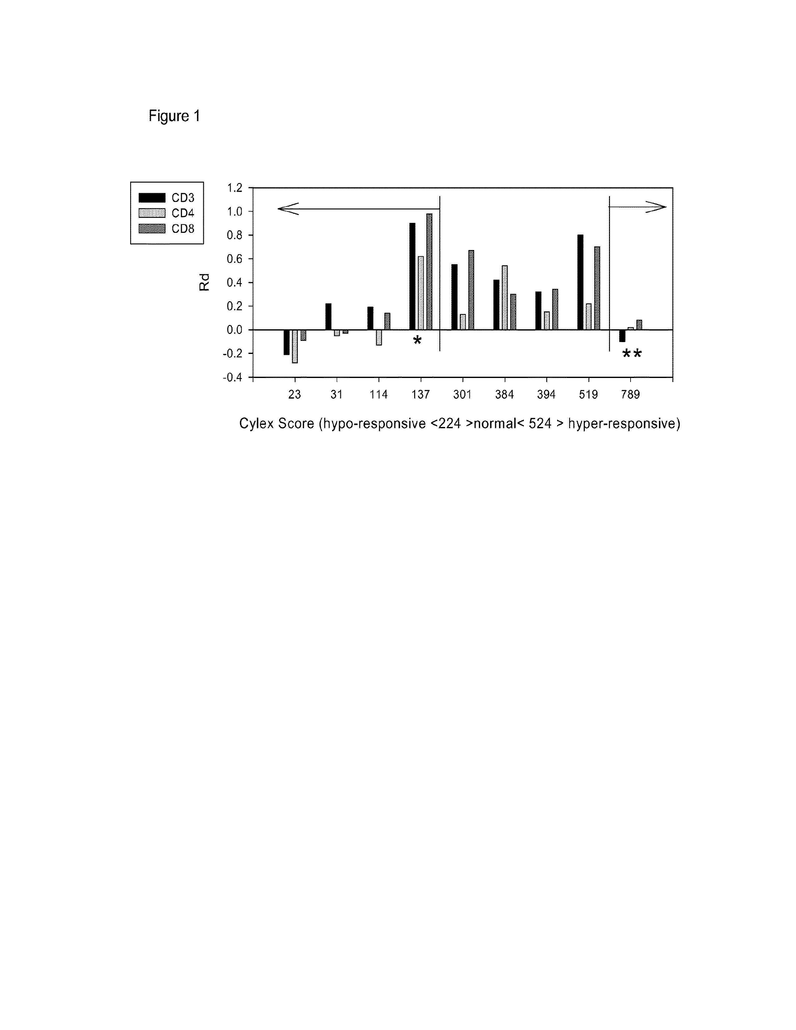
[0058]We measured the degree to which NFκB translocation in peripheral T cells was impaired by immune-suppressive therapy using a commercially available imaging cell flow cytometer (Amnis Corporation, Seattle, Wash.).

[0059]Peripheral blood cells from 9 transplant recipients were isolated, stimulated in culture with PMA / ionomycin (30 min), stained for T cell surface markers and NFκB (p65) and the relative amount of nuclear NFκB in resting and activated CD3, CD4 and CD8 positive T-cell subsets was compared. Results were then correlated with clinical response (stable graft function, infections and rejections) and ImmuKnow assay results. The assay correlated well with results obtained in parallel using the commercially available ImmuKnow product according to manufacturer's instructions, (FIG. 1) but there were 2 major discrepancies. In one patient (** in FIG. 1), the ImmuKnow assay levels indicated heightened immunity yet the patient suffered from major viral infections. In this patient...

example 2

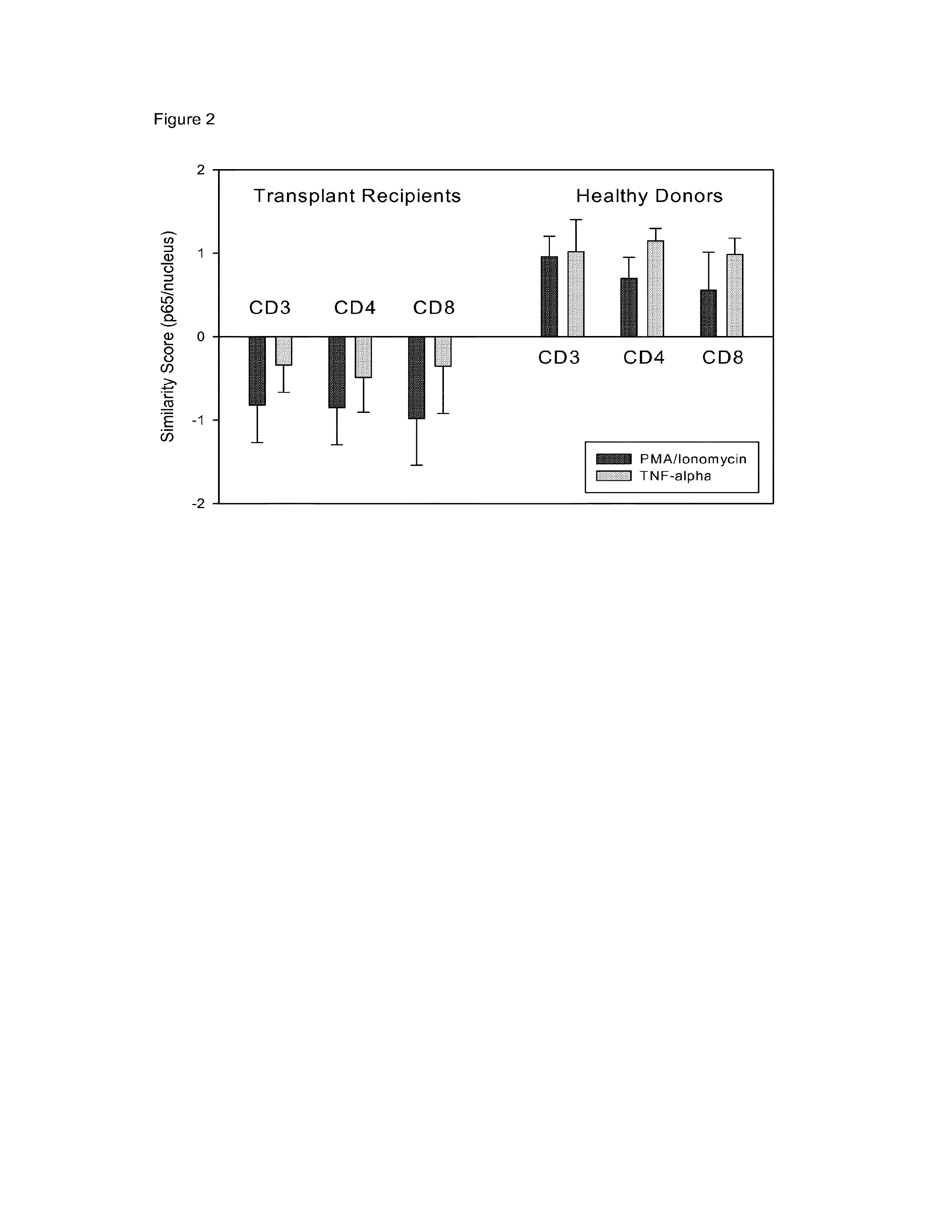
[0060]The assay described in Example 1 was modified to perform the stimulation and cell surface labeling in whole blood to enable the method to be performed in the normal environment of the cells and to permit faster performance of the assay. Using this approach, the immune response of 5 transplant patients as compared to 4 normal donors to stimulation to TNFα or PMA / ion was compared. The data depicted in FIG. 2 demonstrate that using a similarity score read-out for nuclear NFκB as a measure for immune response, a striking difference could be observed between the samples from normal donors and samples from transplant recipients undergoing immunosuppressive therapy. Thus, this Example unexpectedly demonstrates that the method of the invention is suitable analysis of the amount of nuclear NFkB using a procedure whereby immune cells are activated in whole blood.

example 3

[0061]The results presented in this Example were performed using a commercially available imaging cell flow cytometer (Amnis Corporation, Seattle, Wash.) as in Example 1. The materials and methods utilized to obtain the results presented in the Example are essentially as follows:

[0062]Protocol to measure the NF-κB and / or NFAT activation potential in immunophenotypically defined cell populations: Obtain peripheral blood sample in sodium heparin container. Keep PBL sample at ambient room temperature. Prepare sufficient number of 15 mL polypropylene tubes (10-9152N, Niagara Scientific, Lancaster, N.Y.) to accommodate the number of variables to be tested. One tube is needed for the unstimulated control, one for the unstimulated cell line control (HL60 30 or Jurkat) and two (1 for HL60 and 1 for patient sample) for each of the stimulation conditions to be tested (e.g. PMA / Ion, TNFα, CD3 / CD28, etc.). Aliquot 500 μl whole blood per tube for the patient samples. For cell line controls, add ...


PUM

PropertyMeasurementUnit
volumeaaaaaaaaaa
volumesaaaaaaaaaa
volumesaaaaaaaaaa

Abstract

Provided is a method for determining immunosuppression in an individual. The method entails testing blood cells for nuclear NFkB and/or nuclear NFAT. The blood cells can be from a sample of blood from an individual. The cells can be contacted with an activating agent to obtain activated cells, and the amount of nuclear NFkB and/or NFAT can be compared to a control. An amount of nuclear NFkB and/or NFAT that is higher than the control is considered to be indicative of insufficient immunosuppression in the individual. An amount of nuclear NFkB and/or NFAT that is lower than the control is considered to be indicative of excessive immunosuppression in the individual. An amount of nuclear NFkB and/or NFAT that is the same as the control is considered to be indicative of an appropriate amount of immunosuppression in the individual.

Description

CROSS REFERENCE TO RELATED APPLICATIONS[0001]This application claims priority to U.S. Provisional Patent Application No. 61 / 553,324, filed on Oct. 31, 2011, and is a continuation in part application of U.S. application Ser. No. 13 / 128,292, filed Sep. 1, 2011, which is a U.S. national phase application of international application no. PCT / US09 / 64010, filed on Nov. 11, 2009, which in turn claims priority to U.S. Provisional Patent Application No. 61 / 113,381, filed on Nov. 11, 2008, the disclosures of each of which are hereby incorporated by reference.STATEMENT REGARDING FEDERALLY SPONSORED RESEARCH[0002]This invention was made with government support under contract no. 1R21CA126667 awarded by the National Institutes of Health. The government has certain rights in the invention.FIELD OF THE INVENTION[0003]The present invention relates generally to determining immunosuppression, and more particularly to determining immunosuppression in an individual by measuring nuclear NFκB and / or Nucl...

Claims


Application Information

Patent Timeline
Patent Type & Authority Applications(United States)
IPC IPC(8): G01N33/68
CPCG01N33/6872G01N2800/245G01N2333/4703
Inventor PANKEWYCZ, OLEH G.LAFTAVI, MARK R.MINDERMAN, HANS
Owner DAVIDOVICH DONNA
Why Eureka
Try Eureka