Processing of interventional radiology images by ECG analysis

a radiology and image processing technology, applied in the field of medical imaging, can solve the problems of inability to achieve, inability to continuously inject contrast agent into patients, and detrimental alignment defect with two superimposed images

Active Publication Date: 2013-07-25
GENERAL ELECTRIC CO
View PDF26 Cites 2 Cited by
  • Summary
  • Abstract
  • Description
  • Claims
  • Application Information

AI Technical Summary

Problems solved by technology

However, it is not possible, for physiological reasons, to continually inject a contrast agent into a patient.
However, any alignment defect with two superimposed images is detrimental.
For example, the practitioner may see the instrument at a position relative to the model that is different from its effective position in relation to the anatomy, which is detrimental to the necessary precision of the practitioner's intervention.
Alignment defects may result from the patient's physiological movements: for example heart beats and breathing.
These movements may make guiding of the instrument more complex since the practitioner only has access to real time images in which the instrument may be shown at an inaccurate point relative to the data provided by the mask.

Method used


Image

  • Processing of interventional radiology images by ECG analysis
  • Processing of interventional radiology images by ECG analysis
  • Processing of interventional radiology images by ECG analysis

Examples


Embodiment Construction

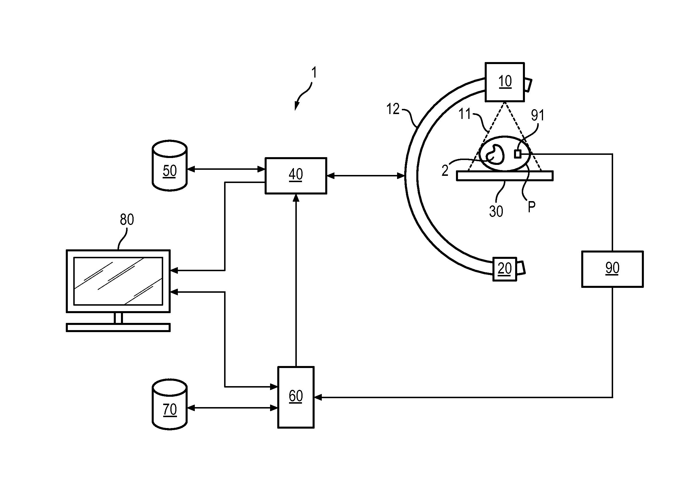
[0019]In all the figures, similar parts carry identical reference numbers.

[0020]In the course of an interventional radiology procedure, a practitioner may move one or more surgical instruments towards a region of interest in a patient by way of the patient's vascular system. The surgical instrument may be a catheter, whether or not equipped with electrodes, a guide wire, or any other instrument known to a person skilled in the art.

[0021]To facilitate moving of the instrument, a medical imaging system allows for the display of the region of interest (region to be treated) in real time. By means of this image, the practitioner may optionally visualize the position of the surgical instrument. The image is a mask of the region of interest which is acquired before the actual procedure. This mask may be a 2D image in which clinical relevant data has been acquired through the injection of a contrast agent, or using any other method known to a person skilled in the art.

[0022]It may also be ...


PUM


Abstract

A method of processing images for interventional imaging, wherein a region of interest is visualized, is provided. The method comprises acquiring a series of 2D images of the region of interest in a patient during at least one respiratory phase, acquiring an electrocardiographic signal which is synchronized with the acquisition of the series of 2D images, processing the electrocardiographic signal to estimate at least one deformation phase of the region of interest induced by the patient's respiratory movement, and registering the different successive 2D images in relation to the estimated deformation phase.

Description

BACKGROUND OF THE INVENTION[0001]Embodiments of the invention concern the field of medical imaging, and in particular, the processing of images in interventional radiology (fluoroscopic images). More specifically it concerns a method and a system with which it is possible in real time to display a region of interest in a patient, in two or three dimensions, in which a surgical instrument can be inserted. Interventional radiology consists of a practitioner guiding and deploying one or more surgical instruments inside a patient's vascular system with the assistance of a medical imaging system.[0002]The medical imaging system allows the acquisition, processing and real-time display of two-dimensional images (2D) showing the patient's vascular system and the surgical instrument(s). These images enable the practitioner to guide the instrument within the vascular system.[0003]The acquisition of these images requires emitting of a low X-ray dose. The vessels are visible therein by means of...

Claims


Application Information

Patent Timeline
Patent Type & Authority Applications(United States)
IPC IPC(8): A61B6/00A61M25/09A61B6/02
CPCA61B6/486A61B6/504A61B6/541A61B6/5288A61B6/485A61B6/548A61B6/54A61B6/4208A61B6/461A61M25/09A61B19/5244A61B6/02A61B34/20A61B6/4441
Inventor CAZALAS, MAXIMEVAILLANT, REGIS
Owner GENERAL ELECTRIC CO
Why Eureka
Try Eureka