
Method and apparatus for intervention imaging in magnetic resonance tomography

a magnetic resonance tomography and intervention imaging technology, applied in the field of magnetic resonance tomography, can solve the problems of weakening or obliterating the magnetic resonance signal, difficult to make the already-bright blood in the mrt image even brighter, and limiting the use of contrast agents to significantly expand the limits of native mr imaging, so as to improve the contrast agent-supported interventional imaging

Inactive Publication Date: 2009-07-07
SIEMENS AG
View PDF11 Cites 0 Cited by
  • Summary
  • Abstract
  • Description
  • Claims
  • Application Information

AI Technical Summary

Benefits of technology

"The present invention provides a method for improving contrast agent-supported interventional imaging in magnetic resonance tomography. This is achieved by preparing the contrast agent liquid in a way that leads to a strong signal representation in the vessel system of a patient to be examined. This can be done by emitting one or more saturation pulses or an inversion pulse into the stationary tissue or by conducting the contrast agent liquid through a static magnetic gradient field and using continuous RF irradiation. The invention also includes a device suitable for implementing the inventive method. The technical effect of the invention is to improve the accuracy and reliability of interventional imaging in magnetic resonance tomography."

Problems solved by technology

Nevertheless, particularly in angiographic exposures (i.e. exposures of the blood vessels in the human body, especially in organs supplied (perfused) with blood), the contrasting in native MR imaging sets limits that can be significantly expanded by the use of contrast agents.
However, it is difficult to make the already-bright blood in the MRT image even brighter.
According to the prior art, this ensues, for example, by injection of magnetic liquids that induce susceptibility artifacts and that significantly weaken or obliterate the magnetic resonance signal.
This method is limited, however, to only a few injection measurements because the recommended highest dose of these substances is very low.
CO2 is in fact a suitable contrast agent that has already been used for a long time in interventional radiology; nevertheless, many users (doctors) balk at injecting a gas into blood vessels.
A disadvantage is also that this method can be applied only below the diaphragm.
The application of CO2 at the heart or at cerebral vessels is forbidden due to the embolism risk that exists.
Disadvantages of this method are the short T1 time of the blood (approximately 1500 ms, approximately 100 ms after contrast agent administration) as well as the limitation to applications with sufficient blood flow.

Method used


Image

  • Method and apparatus for intervention imaging in magnetic resonance tomography
  • Method and apparatus for intervention imaging in magnetic resonance tomography
  • Method and apparatus for intervention imaging in magnetic resonance tomography

Examples


Embodiment Construction

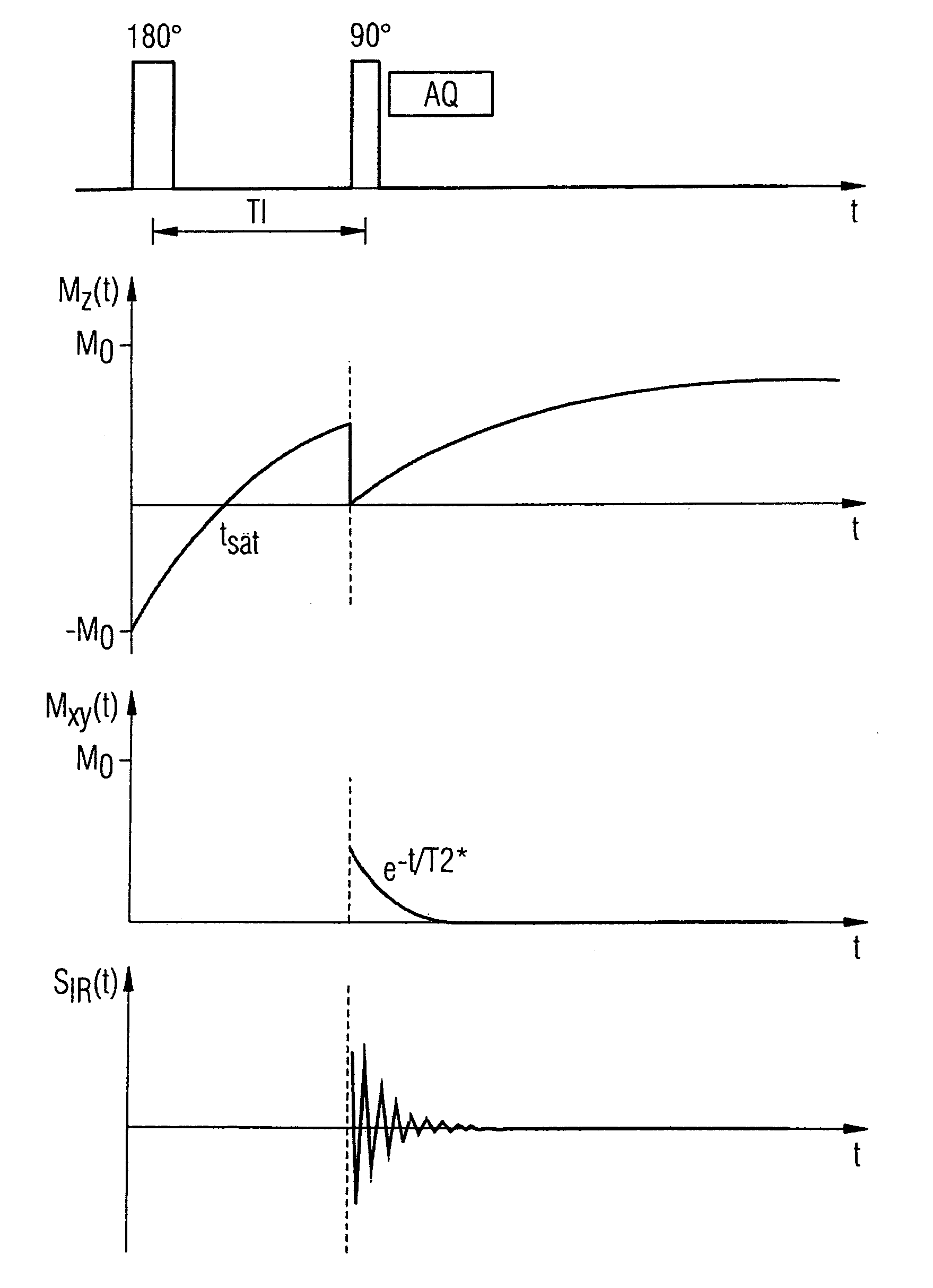
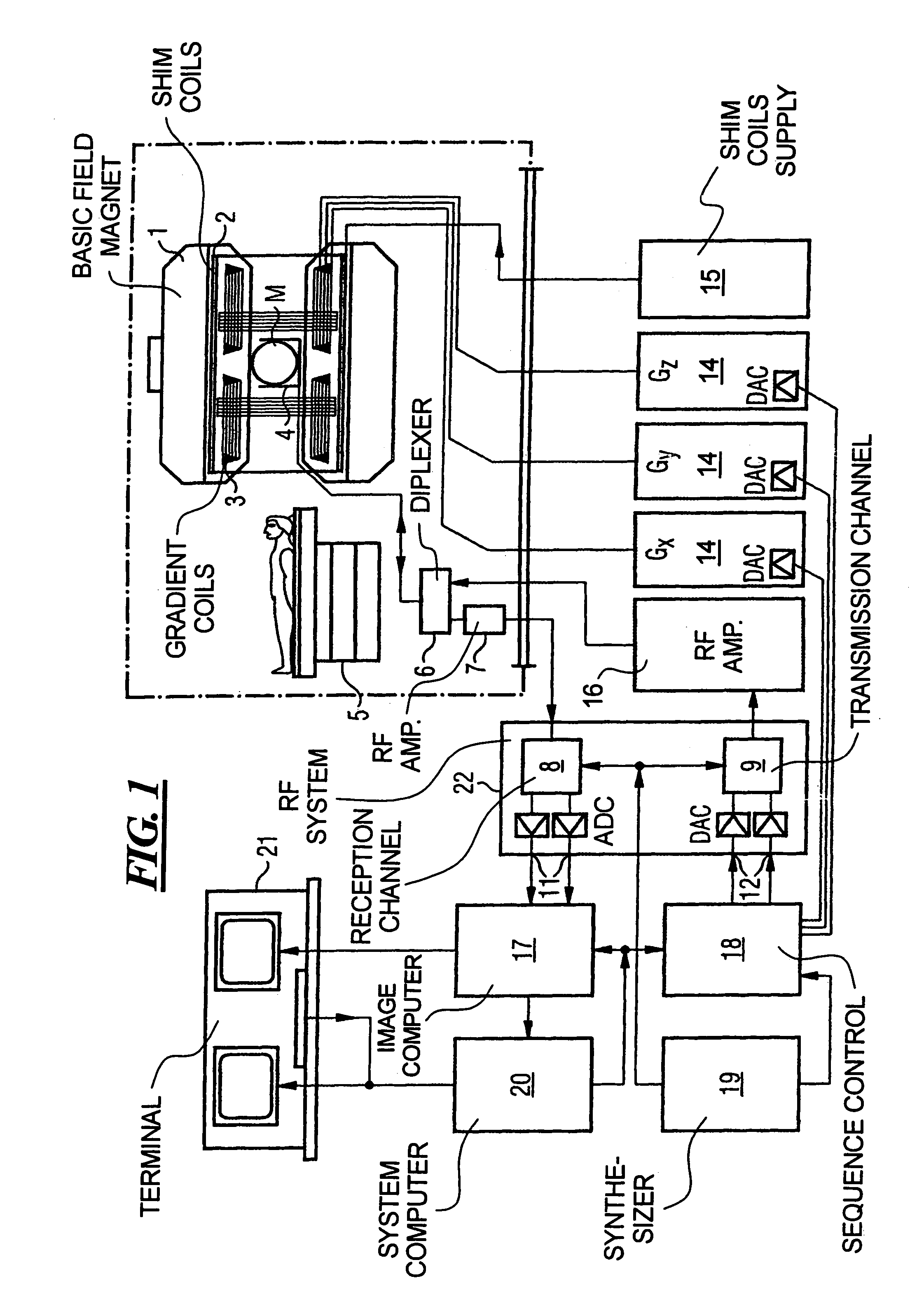
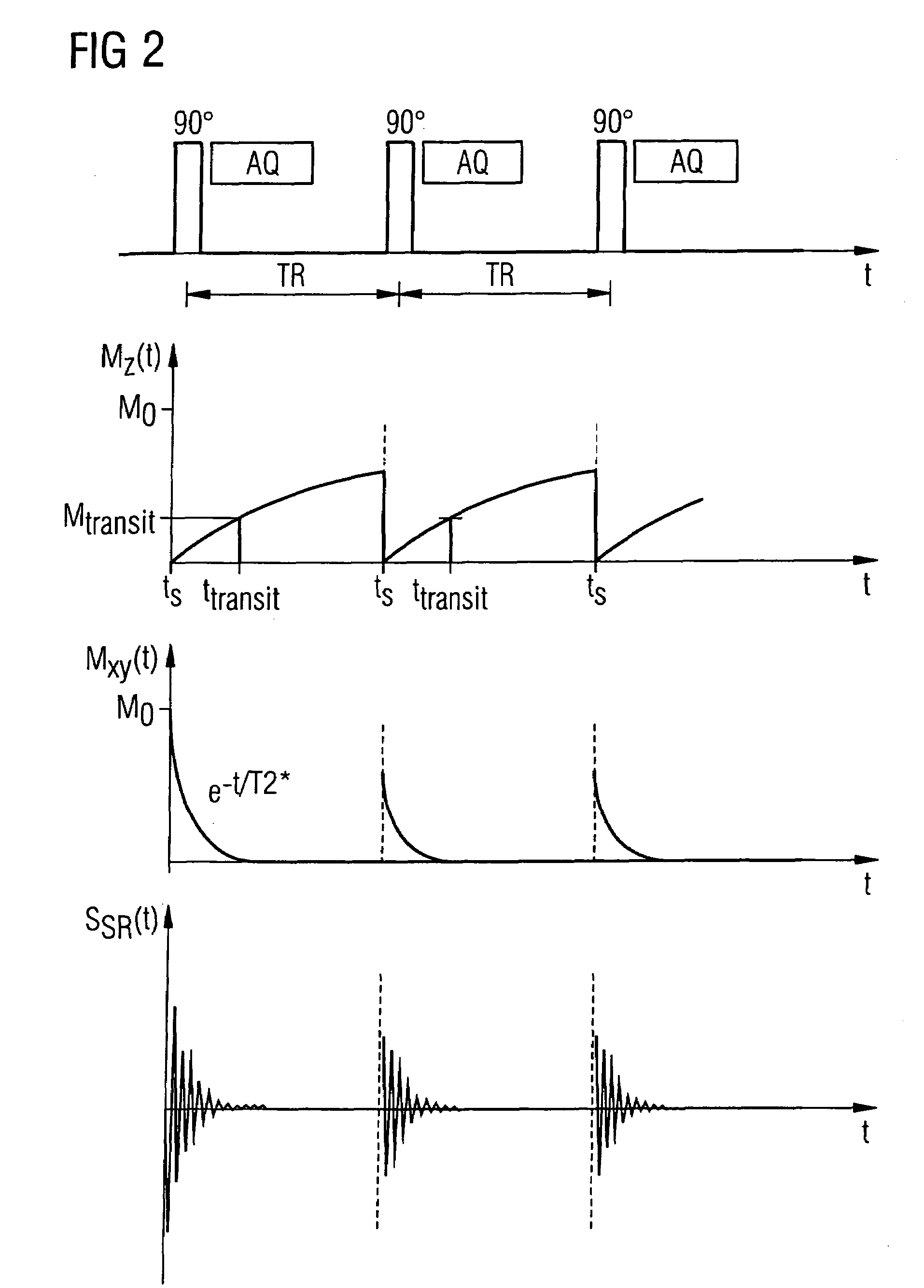
[0031]FIG. 1 is a block diagram of a magnetic resonance tomography apparatus with which an interventional imaging is possible according to the present invention. The component of the magnetic resonance tomography apparatus correspond to the design of a conventional tomography apparatus with the exceptions described below. A basic field magnet 1 generates a temporally constant strong magnetic field for polarization or alignment of the nuclear spins in the examination region of a subject such as, for example, of a part of a human body to be examined. The high homogeneity of the basic magnetic field necessary for the magnetic resonance scan is defined in a spherical measurement volume M in which the parts of the human body to be examined are introduced. To satisfy the homogeneity requirements, and in particular for elimination of temporally non-varying influences, what are known as shim plates made from ferromagnetic material are mounted at a suitable location. Temporally variable infl...


PUM


Abstract

In a method and apparatus for interventional imaging in magnetic resonance tomography, a contrast agent liquid is prepared by saturation or excitation of the nuclear spins therein, such that it shows with a poor signal in the vessel system of a patient to be examined after injection, or the stationary tissue is prepared by saturation or excitation of the nuclear spins therein such that the contrast agent liquid shows with strong signal (compared to the tissue) in the vessel system of a patient to be examined after injection.

Description

BACKGROUND OF THE INVENTION[0001]1. Field of the Invention[0002]The present invention generally concerns magnetic resonance tomography (MRT) as employed in medicine for examination of patients. The present invention in particular concerns a method for improved interventional imaging in MRT using contrast agent liquids.[0003]2. Description of the Prior Art[0004]MRT is based on the physical phenomenon of nuclear magnetic resonance and has been successfully used as an imaging method for over 20 years in medicine and biophysics. In this examination modality, the subject is exposed to a strong, constant magnetic field. The nuclear spins of the atoms in the subject, which were previously randomly oriented, thereby align. Radio-frequency energy can now excite these “ordered” nuclear spins to a specific oscillation. In MRT, this oscillation generates the actual measurement signal that is acquired by appropriate reception coils. By the use of inhomogeneous magnetic fields generated by gradie...

Claims


Application Information

Patent Timeline
Patent Type & Authority Patents(United States)
IPC IPC(8): A61B5/055A61B5/05A61B5/06G01R33/28G01R33/563
CPCA61B5/055A61B5/411A61B5/06
Inventor SPEIER, PETERWACKER, FRANK
Owner SIEMENS AG
Learn More