

Non-radioactive agents for neuroblastoma imaging
a neuroblastoma and imaging technology, applied in the field of imaging agents, can solve the problems of increasing the risk of secondary cancer, increasing the risk of neuroblastoma secondary cancer, and increasing the toxicity of therapy
- Summary
- Abstract
- Description
- Claims
- Application Information
AI Technical Summary
Benefits of technology
Problems solved by technology
Method used
Image


Examples
example 1
Methods and Materials
[0070]Cell lines. Human neuroblastoma cancer cell lines NB1691.1uc and SYFY.luc were cultured in Dulbecco's Modified Eagle's Medium supplemented with high glucose and F12 nutrient (DMEM / F12, Invitrogen, Carlsbad, Calif.) with 10% fetal bovine serum (Hyclone, Logan, Utah) in a humidified incubator maintained at 37° C. with 5% CO2.
[0071]Tumor xenografts: Four- to six-week-old female nude mice (18-22 g) (Taconic, Hudson, N.Y.) were housed and fed with sterilized pellet chow and sterilized water. Animals were maintained in a pathogen-free mouse colony. Tumor cells near confluence were harvested by incubation with 0.05% trypsin-EDTA. Cells were pelleted by centrifugation at 130×g for 5 min and resuspended in sterile phosphate-buffered saline (PBS). Approximately 1 million cells were implanted subcutaneously into the hind leg region of the mice. A total of 15 mice was used in this study.
[0072]Confocal microscope imaging. Cells were harvested from culture and incubated...
example 2
Synthesis of W765-BG
[0076]The reaction scheme and structure of W765-BG is shown in Scheme 1. The synthesis procedure was carried out in four steps. Compound 1 (3-aminobenzyl alcohol) was reacted with Boc-Gly-OSu under basic conditions to form 3-(Boc-Gly-amino)benzyl alcohol (Compound 2). By means of the Mitzunobu protocol (Dodd and Kozikowski, 1994), compound 2 was converted to 1,3-bis(tert-butyloxycarbonyl)-2-(3-(Boc-Gly-amino)benzyl)guanidine (compound 3) by treatment with N,N-bis-Boc-guandine, triphenylphosphine, and diidsopropyl azo-dicarboxylate. Three Boc-protecting groups on compound 3 were removed under acidic conditions, resulting in compound 4. This compound was conjugated to IRdye800cw through the α-amino functional group of glycine to result in the NIR optical imaging agent W765-BG.
[0077]Synthesis of 3-(Boc-Gly-amino)benzyl alcohol (Boc-Gly-NH-Bn-OH) (Compound 2): 3-aminobenzyl alcohol (123 mg, 1 mmol) and Boc-Gly-OSu (272 mg, 1 mmol) were dissolved in 5 mL of DMF. DIPEA...
example 3
Imaging using W765-BG
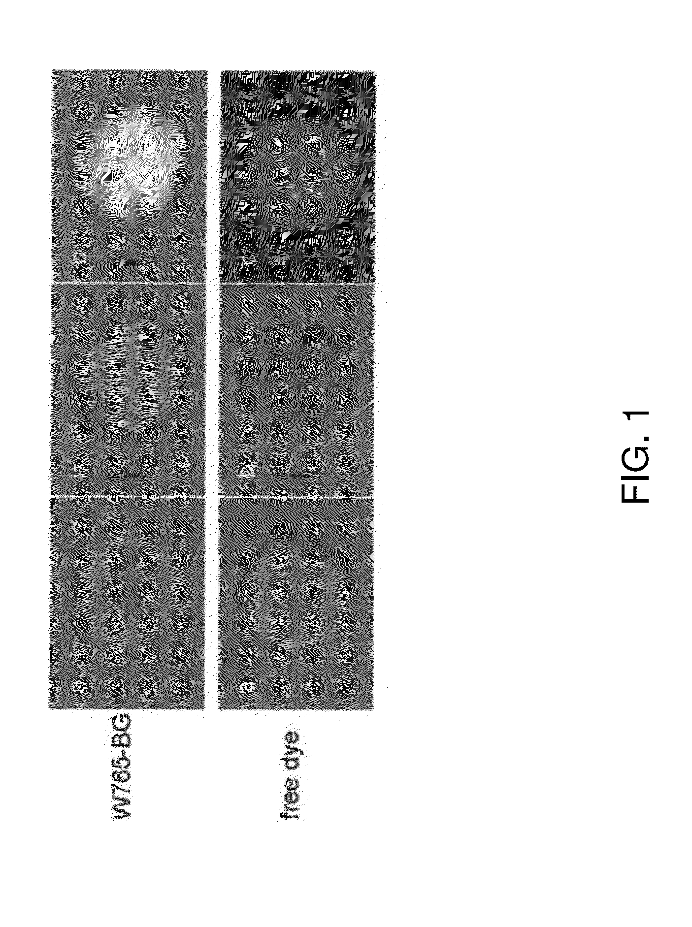
[0081]Imaging of W765-BG in neuroblastoma cells. To evaluate W765-BG as a potential specific molecule, its ability to uptakes by neuroblastoma cells was examined in vitro by NIR confocal microscopy. NPG.Luc cells were treated with W765-BG or free dye overnight at 37° C., after which they were washed, fixed, and visualized by confocal microscopy. W765-BG was detected inside the cells (FIG. 1, top panel), while there was almost no detectable signal in cells treated with the free dye (IRDye800CW carboxylate) alone (FIG. 1, bottom panel). This data confirms that W765-BG is taken up by neuroblastoma cells. The merged confocal images clearly demonstrate that W765-BG is located inside the cell and in the nuclear compartment.
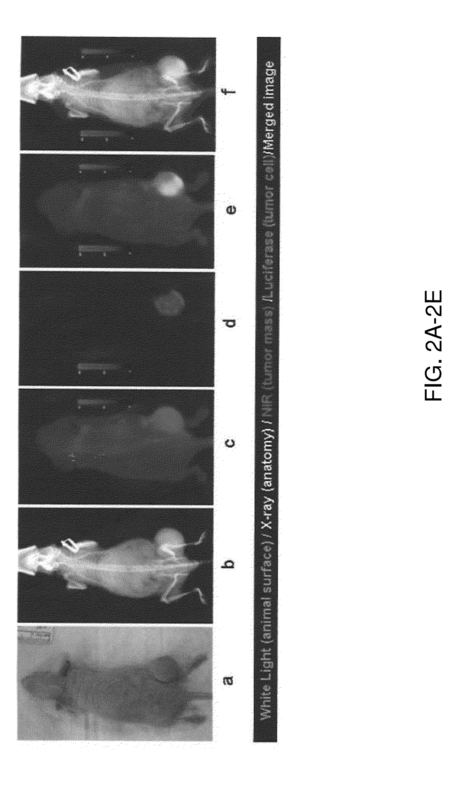
[0082]Imaging of W765-BG in human tumor xenografts in mice. To further demonstrate the feasibility of imaging neuroblastomas using W765-BG, luciferase-positive tumor cells (NGP.Luc) were implanted subcutaneously into the hind region of mice. Tumor-b...
PUM
 Abstract
Description
Claims
Application Information