Diagnostic imaging apparatus and method

a diagnostic imaging and imaging apparatus technology, applied in the field of diagnostic imaging apparatus and method, can solve the problems of difficult determination, subjective interpretation of the shape of the lesion, and shape interpreters may not be able to adequately interpret medical images with significant speckle nois

Inactive Publication Date: 2013-09-19
SAMSUNG ELECTRONICS CO LTD
View PDF6 Cites 16 Cited by
  • Summary
  • Abstract
  • Description
  • Claims
  • Application Information

AI Technical Summary

Problems solved by technology

Internal echoes and rear echo shades are additional factors, but may be difficult to determine, especially in sonograms with significant noise or speckles.
However, the interpretation of the shape of a lesion may be subjective and may thus vary depending on the radiologist.
However, shape interpreters may not be able to adequately interpret medical images having significant speckle noise, such as sonograms.
In addition, since shape interpreters are limited to the morphological distance and angle between shapes, it may be difficult to properly interpret highly irregular lesion shapes.

Method used


Image

  • Diagnostic imaging apparatus and method
  • Diagnostic imaging apparatus and method
  • Diagnostic imaging apparatus and method

Examples


Embodiment Construction

[0036]The following description is provided to assist the reader in gaining a comprehensive understanding of the methods, apparatuses, and / or systems described herein. Accordingly, various changes, modifications, and equivalents of the methods, apparatuses, and / or systems described herein may be suggested to those of ordinary skill in the art. Also, descriptions of well-known functions and constructions may be omitted for increased clarity and conciseness.

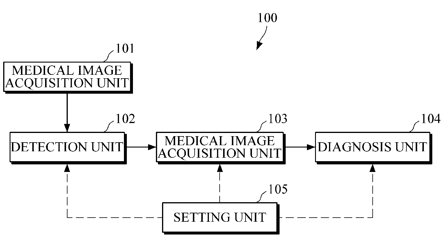
[0037]FIG. 1 is a diagram illustrating an example of a diagnostic imaging apparatus.

[0038]Referring to the example illustrated in FIG. 1, a diagnostic imaging apparatus 100 includes a medical image acquisition unit 101, a detection unit 102, an interpretation unit 103, a diagnosis unit 104, and a setting unit 105.

[0039]The medical image acquisition unit 101 acquires a medical image. In an example, the acquired medical image is one or more of the group consisting of a radiograph, a sonogram, a magnetic resonance imaging (MRI) image,...


PUM


Abstract

A diagnostic imaging apparatus and method are provided. The diagnostic imaging apparatus includes a detection unit configured to detect a lesion from a medical image, an interpretation unit configured to acquire a shape feature value by interpreting a shape of the detected lesion in a frequency domain, and a diagnosis unit configured to determine whether the detected lesion is benign or malignant based on the acquired shape feature value.

Description

CROSS-REFERENCE TO RELATED APPLICATIONS[0001]This application claims the benefit under 35 U.S.C. §119(a) of Korean Patent Application No. 10-2012-0026152, filed on Mar. 14, 2012, in the Korean Intellectual Property Office, the entire disclosure of which is incorporated herein by reference for all purposes.BACKGROUND[0002]1. Field[0003]The following description relates to a diagnostic imaging apparatus and method.[0004]2. Description of Related Art[0005]Breast sonography is performed to detect masses or lesions that may not be detected by mammography, thereby reducing the need to perform biopsies that may prove to be unnecessary. A mass or lesion may be defined as a suspicious lump of tissues that differs from its surroundings. Women over the age of forty may benefit from breast sonography when mammography detects suspicious findings. Further, breast sonography is recommended for young, skinny women or pregnant women after mammography. The sensitivity of mammography is relatively low...

Claims


Application Information

Patent Timeline
Patent Type & Authority Applications(United States)
IPC IPC(8): A61B6/00A61B5/00A61B6/03A61B5/055A61B8/08
CPCA61B8/5223A61B6/502A61B5/055A61B8/085A61B6/5217A61B5/7257G06T2207/30068G06T7/0014G06T2207/10132A61B6/032G16H50/30A61B6/00A61B8/08G16Z99/00G06F18/2413G06T7/0012
Inventor LEE, JONG-HASEONG, YEONG-KYEONG
Owner SAMSUNG ELECTRONICS CO LTD
Why Eureka
Try Eureka