
Endoscope attachment and method

a technology of endoscope and endoscope, which is applied in the field of endoscope attachment and method, can solve the problems of limited size of catheters, difficult procedures, and difficult medical tube placement procedures

Inactive Publication Date: 2014-03-13
LUCENT MEDICAL SYST
View PDF3 Cites 10 Cited by
  • Summary
  • Abstract
  • Description
  • Claims
  • Application Information

AI Technical Summary

Benefits of technology

The patent describes an endoscopic device with a retention channel for holding a medical device in place. The device has a separation mechanism between the medical device and the retention channel, which can be released by pulling a cable assembly. The device also has a tubular body with a secondary channel for receiving the cable assembly. The invention allows for easy placement of a medical device in a patient's body with an endoscope. The technical effects of the invention include improved placement of medical devices and ease of use for medical professionals.

Problems solved by technology

The procedure can be difficult.
In many cases, unless the target site can be visualized directly or unless there is a pathway that is reasonably direct and confined, the medical tube placement procedure can be challenging.
For example, the size of the catheter that can be placed is limited by the strict size dimensions of the endoscope's tool channel.
If the endoscope and associated tool channel diameter is small, adequate space for mechanical clearance of the catheter will not be available.
The snare approach reduces the size concerns that arise when a catheter is placed in an endoscopic tool channel and the endoscope is withdrawn over the catheter, but the snare approach increases the overall complexity of the procedure.
For example, in many cases, the addition of the snare tool, which has actuation and control independent of the endoscope control, adds the possibility that the tube may be dropped during transit.
Moreover, the medical device that rides alongside the endoscope increases the volume and surface area of foreign material passed into the patient.

Method used


Image

  • Endoscope attachment and method
  • Endoscope attachment and method
  • Endoscope attachment and method

Examples


Embodiment Construction

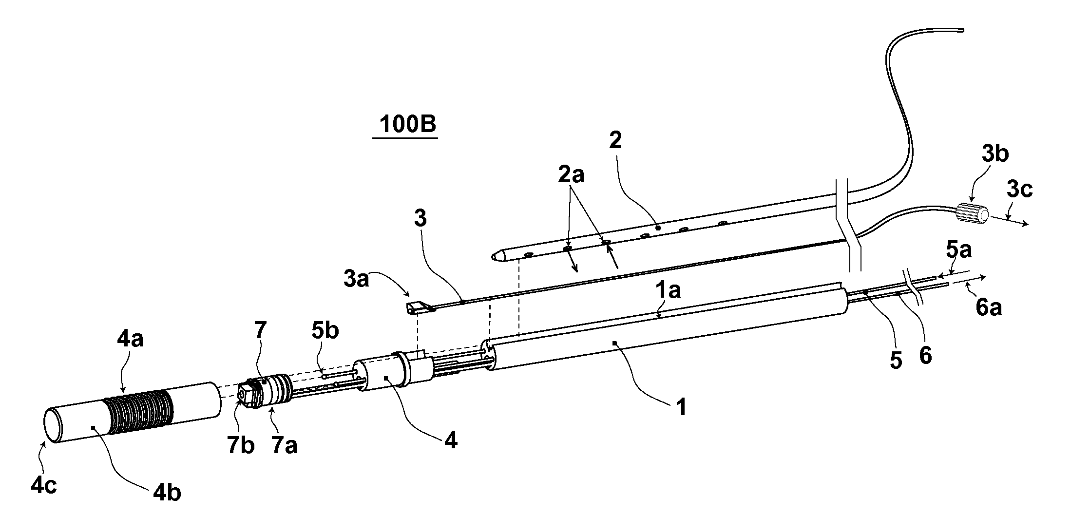
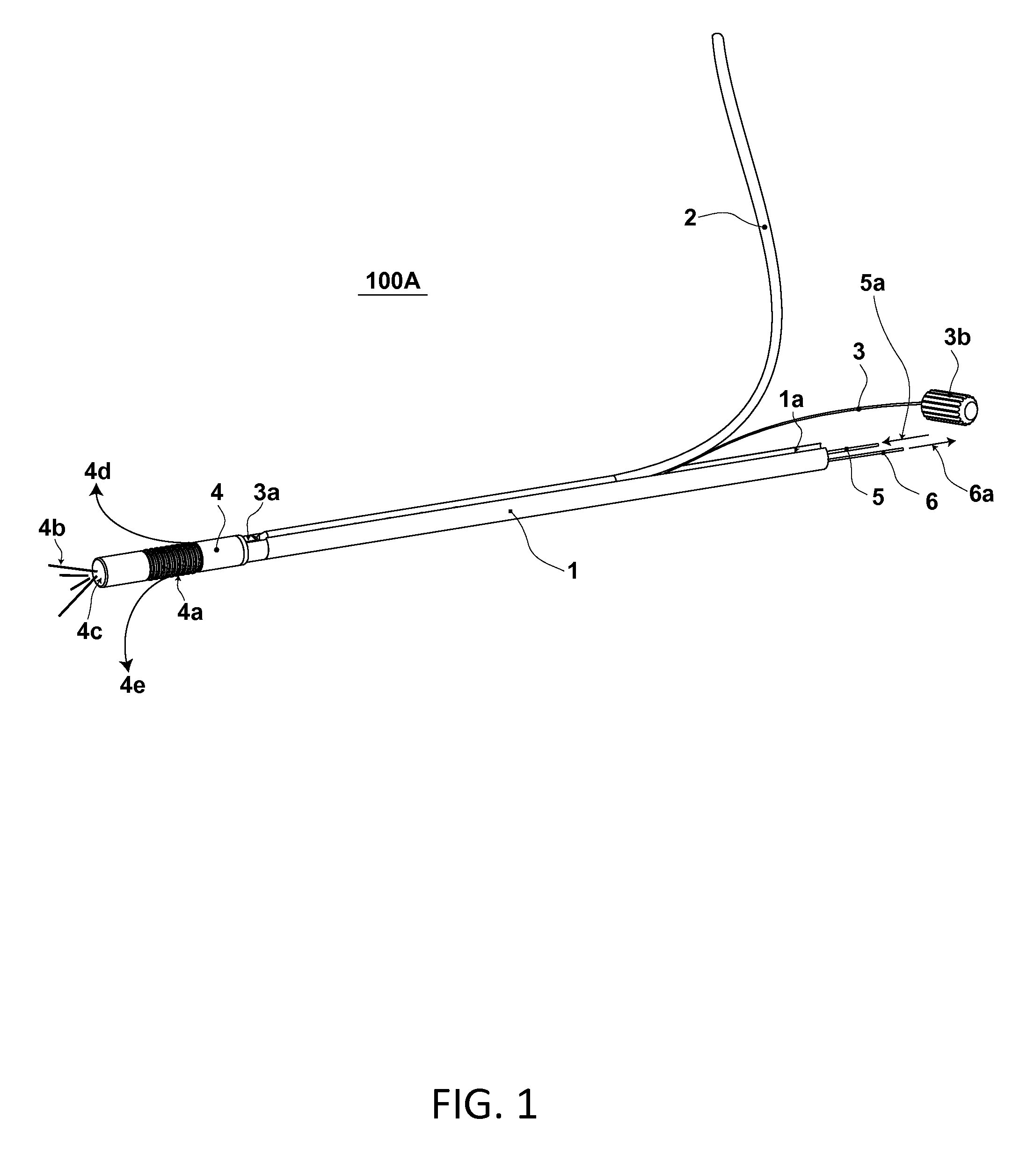
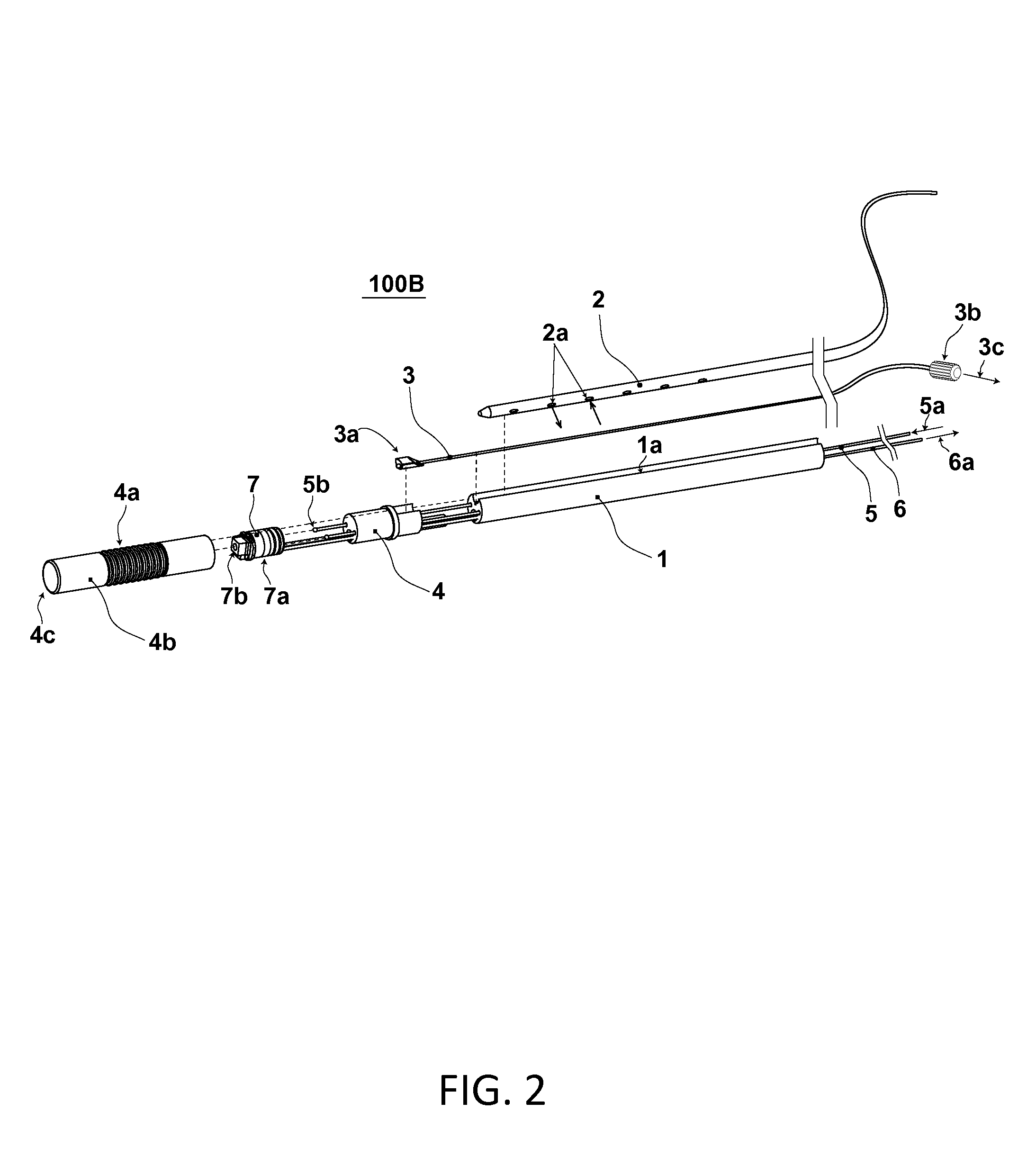
[0034]Embodiments in the present disclosure describe devices and methods of delivering independent, external catheter tubes and other medical devices to a location in a patient's body. Several embodiments are described herein with respect to using direct endoscopic visualization, but the concepts and devices described and illustrated are not so limited. In fact, in addition or as an alternative to endoscopes (e.g., bronchoscopes, colonoscopes, gastrointestinal endoscopes, nasopharyngoscopes, sigmoidoscopes, and the like), other non-endoscopic medical device delivery tools can be used. Accordingly, an endoscope or some other medical device delivery tool can be used in cooperation with the novel attachment / detachment concepts described herein.

[0035]Once a medical device (e.g., a catheter) is put into a desirable position in the patient's body, a method to release the medical device from the medical device delivery tool is executed. The method permits the medical device delivery tool (...


PUM


Abstract

An endoscopic device has a tubular body with a retention channel substantially extending axially along the tube body. A medical tube is removably arranged in the retention channel and held in place by friction. A cable assembly is arranged between the medical tube and the surface of the retention channel. The cable assembly includes a shaped end fixture that is drawn between the medical tube and the retention channel when the cable is pulled, thereby releasing the medical tube from the tube body.

Description

CROSS-REFERENCE TO RELATED APPLICATION[0001]This application claims the benefit under 35 U.S.C. §119(e) of U.S. Provisional Patent Application No. 61 / 699,200 filed Sep. 10, 2012, where this provisional application is incorporated herein by reference in its entirety.BACKGROUND[0002]1. Technical Field[0003]The present disclosure generally relates to an attachment for an endoscope type instrument and method to use the attachment. More particularly, but not exclusively, the present disclosure relates to placing a device such as a medical tube in the body of a patient using an apparatus integrated with or attached to an endoscopic-type instrument.[0004]2. Description of the Related Art[0005]In many medical procedures, it is often beneficial to place a medical device into the body of a patient. For example, one common medical practice is placement of a medical tube, which is carried out using an endoscope (e.g., bronchoscope, colonoscope, gastrointestinal endoscope, nasopharyngoscope, sig...

Claims


Application Information

Patent Timeline
IPC IPC(8): A61B1/018
CPCA61B1/018A61B1/00073A61B1/0014
Inventor VINCENT, STEVEKING, CURTIS S.GOLDEN, ROBERT N.SILVERSTEIN, FRED E.
Owner LUCENT MEDICAL SYST
Learn More