Antibody-based arrays for detecting multiple signal transducers in rare circulating cells
a signal transducer and array technology, applied in the field of antibodies for detecting multiple signal transducers in rare circulating cells, can solve the problems that biopsy from a single site might not represent the heterogeneity of a tumor population, and the molecular analysis of tumor samples is even more difficul
- Summary
- Abstract
- Description
- Claims
- Application Information
AI Technical Summary
Benefits of technology
Problems solved by technology
Method used
Image
Examples
example 1
Isolation, Stimulation, and Lysis of Circulating Cells
[0195]Circulating cells of a solid tumor comprise cells that have either metastasized or micrometastasized from a solid tumor and include circulating tumor cells (CTCs), cancer stem cells (CSCs), and / or cells that are migrating to the tumor (e.g., circulating endothelial progenitor cells (CEPCs), circulating endothelial cells (CECs), circulating pro-angiogenic myeloid cells, circulating dendritic cells, etc.). Patient samples containing circulating cells can be obtained from any accessible biological fluid (e.g., blood, urine, nipple aspirate, lymph, saliva, fine needle aspirate, etc.). The circulating cells can be isolated from a patient sample using one or more separation methods such as, for example, immunomagnetic separation (see, e.g., Racila et al., Proc. Natl. Acad. Sci. USA, 95:4589-4594 (1998); Bilkenroth et al., Int. J. Cancer, 92:577-582 (2001)), microfluidic separation (see, e.g., Mohamed et al., IEEE Trans. Nanobiosc...
example 2
Single Cell Detection Using a Single Detector Sandwich ELISA with Tyramide Signal Amplification
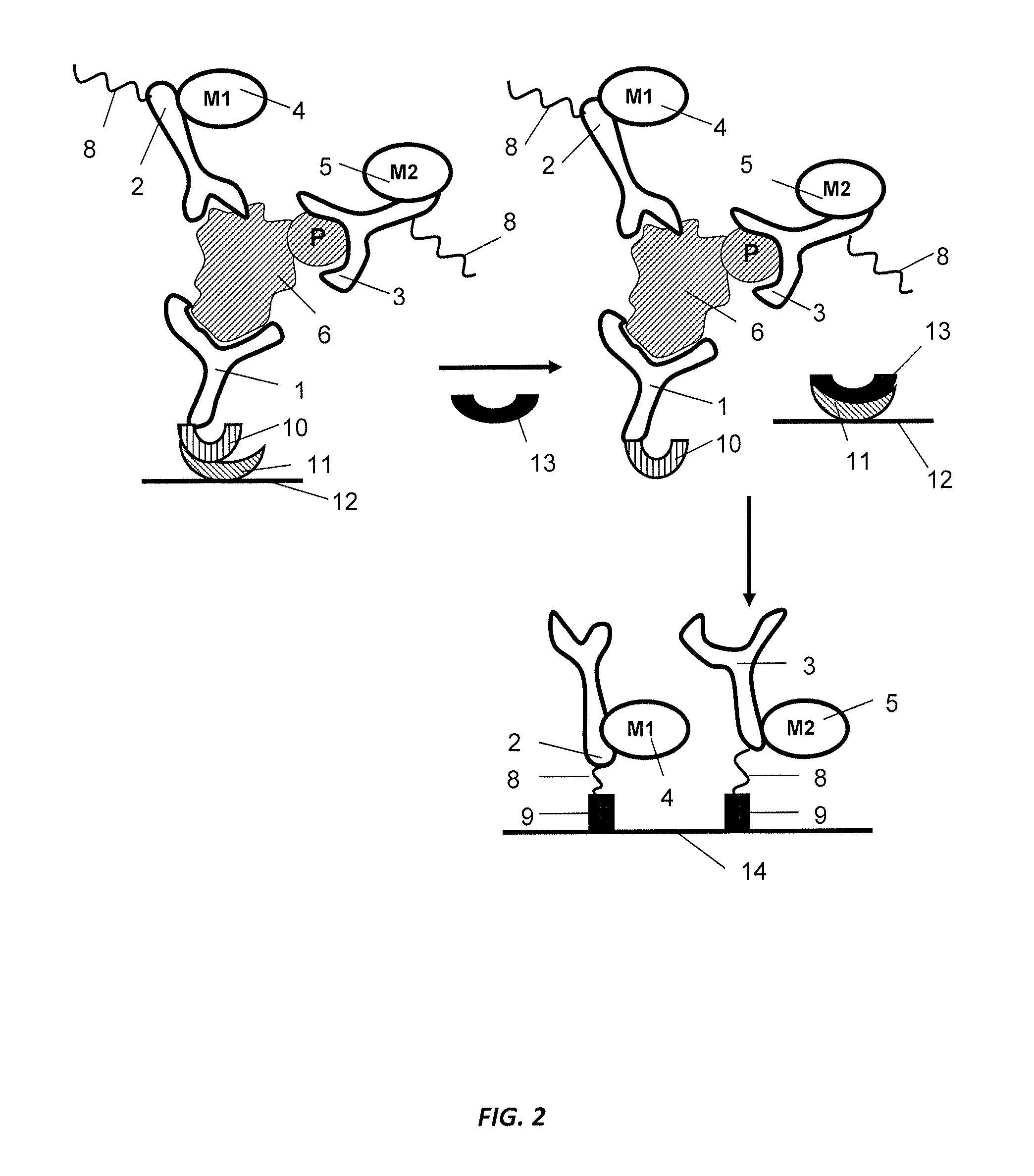
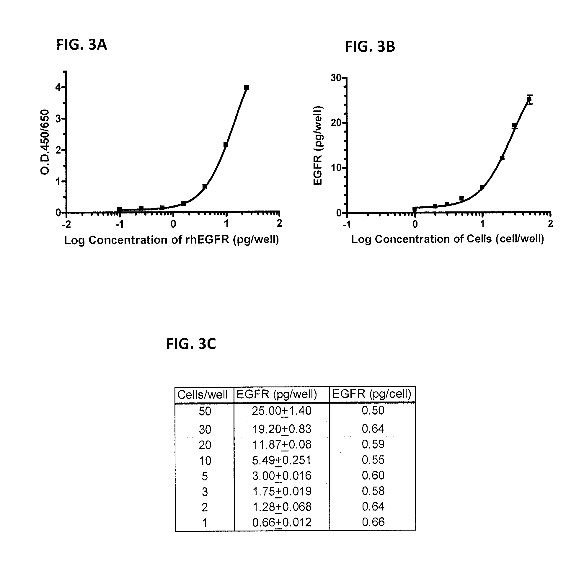
[0320]This example illustrates a multiplex, high-throughput, single detector sandwich ELISA having superior dynamic range that is suitable for analyzing the activation states of signal transduction molecules in rare circulating cells:[0321]1) A 96-well microtiter plate was coated with capture antibody overnight at 4° C.[0322]2) The plate was blocked with 2% BSA / TBS-Tween for 1 hour the next day.[0323]3) After washing with TBS-Tween, the cell lysate or recombinant protein was added at serial dilution and incubated for 2 hours at room temperature.[0324]4) The plate was washed 4 times with TBS-Tween and then incubated with a biotin-labeled detection antibody for two hours at room temperature.[0325]5) After incubation with the detection antibody, the plate was washed four times with TBS-Tween and then incubated with streptavidin-labeled horseradish peroxidase (SA-HRP) for 1 hour at room temper...
example 3
Single Cell Detection Using a Single Detector Microarray ELISA with Tyramide Signal Amplification
[0334]This example illustrates a multiplex, high-throughput, single detector microarray sandwich ELISA having superior dynamic range that is suitable for analyzing the activation states of signal transduction molecules in rare circulating cells:[0335]1) Capture antibody was printed on a 16-pad FAST slide (Whatman Inc.; Florham Park, N.J.) with a 2-fold serial dilution.[0336]2) After drying overnight, the slide was blocked with Whatman blocking buffer.[0337]3) 80 μl of cell lysate was added onto each pad with a 10-fold serial dilution. The slide was incubated for two hours at room temperature.[0338]4) After six washes with TBS-Tween, 80 μl of biotin-labeled detection antibody was incubated for two hours at room temperature.[0339]5) After six washes, streptavidin-labeled horseradish peroxidase (SA-HRP) was added and incubated for 1 hour to allow the SA-HRP to bind to the biotin-labeled det...
PUM
 Abstract
Description
Claims
Application Information
 - R&D
- Intellectual Property
- Life Sciences
- Materials
- Tech Scout
- Unparalleled Data Quality
- Higher Quality Content
- 60% Fewer Hallucinations
Browse by: Latest US Patents, China's latest patents, Technical Efficacy Thesaurus, Application Domain, Technology Topic, Popular Technical Reports.
© 2025 PatSnap. All rights reserved.Legal|Privacy policy|Modern Slavery Act Transparency Statement|Sitemap|About US| Contact US: help@patsnap.com


