Secondary imaging endoscopic device

a technology of endoscopic device and imaging camera, which is applied in the field of secondary imaging endoscopic device, can solve the problems of difficult for a practitioner to identify polyps and contact a detected polyp with a surgical tool,

Inactive Publication Date: 2014-11-20
PSIP LLC
View PDF9 Cites 103 Cited by
  • Summary
  • Abstract
  • Description
  • Claims
  • Application Information

AI Technical Summary

Problems solved by technology

Internal body cavities often have irregular geometries and surface properties that may interfere with imaging and accessing tissues of interest.
For example, since the gastrointestinal tract is tortuous and has a convoluted surface that includes many folds and pockets, it may be difficult for a practitioner to identify polyps and to contact a detected polyp with a surgical tool.

Method used


Image

  • Secondary imaging endoscopic device
  • Secondary imaging endoscopic device
  • Secondary imaging endoscopic device

Examples


Embodiment Construction

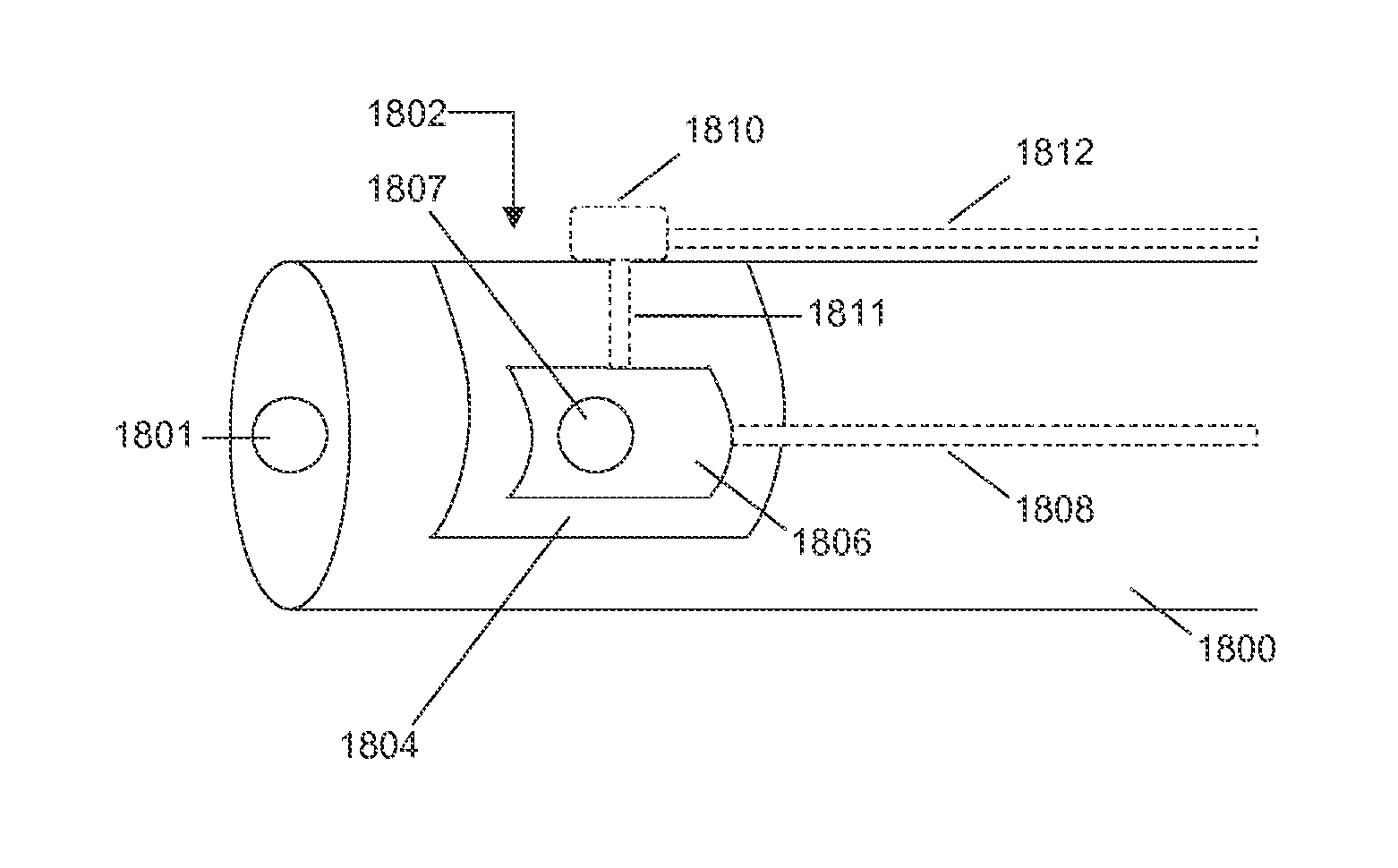
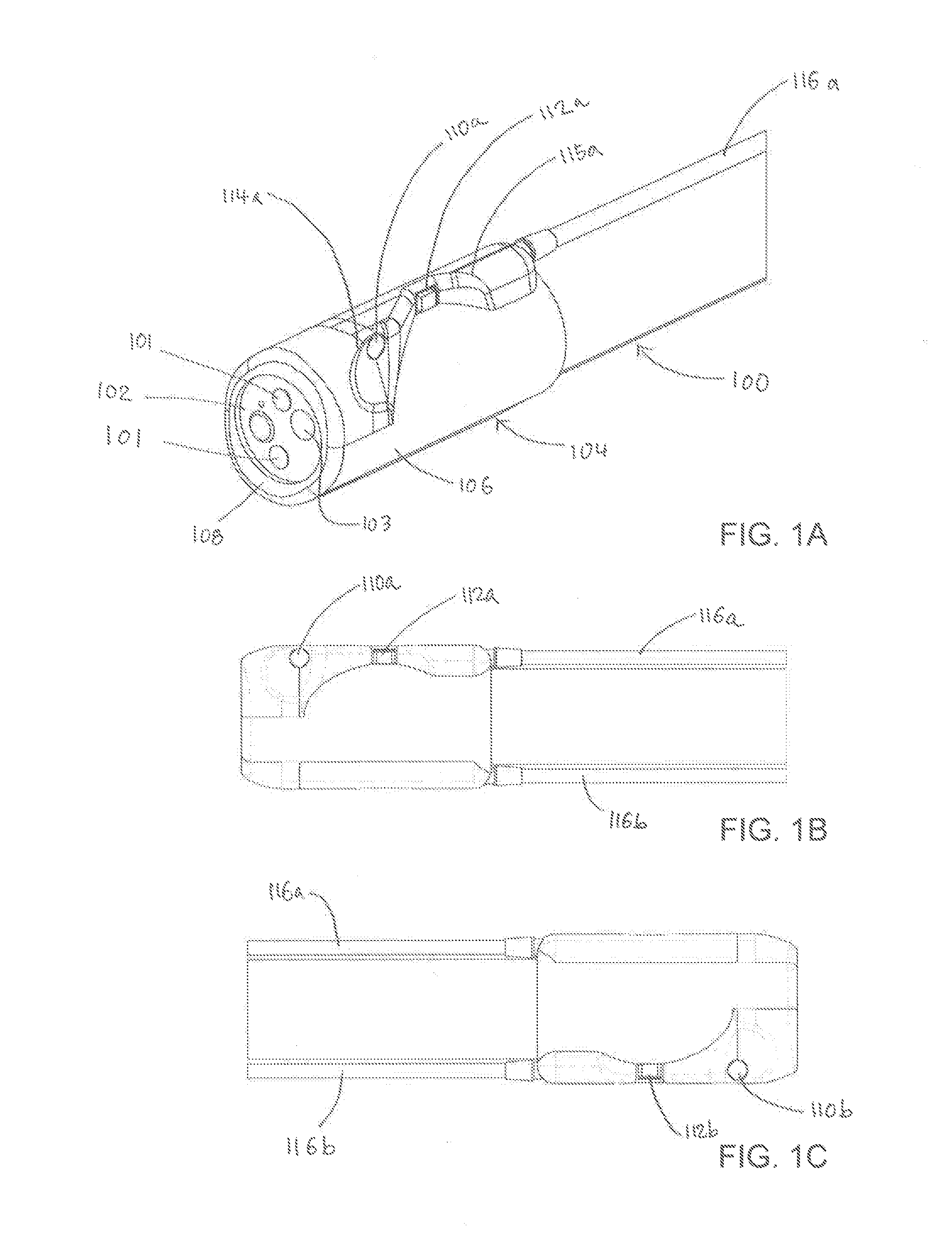
[0033]A secondary imaging endoscopic device may comprise an endoscope attachment member such as a sleeve that is configured to be attached over the distal portion of an endoscope, the sleeve comprising one or more side imaging elements located along the side of the elongated sleeve, and one or more light sources located along the side of the sleeve. Alternatively, a secondary imaging endoscopic device may comprise a detachable endoscope attachment member such as a sleeve or clip and an imaging module attached to the sleeve or clip. The imaging module may comprise one or more side-facing and / or top-facing imaging elements (relative to the front-facing endoscope imaging element) and one or more light sources (e.g., corresponding to each of the imaging elements). Optionally, a secondary imaging endoscopic device may also comprise a fluid delivery module attached to the endoscope attachment member and / or the imaging module, where the fluid delivery module has one or more outlet ports fo...


PUM


Abstract

Described herein are various detachable secondary imaging endoscopic devices that can be used in conjunction with an endoscope to provide additional fields of view so that multiple regions of a body cavity may be imaged simultaneously. In one variation, a secondary imaging endoscopic device comprises an endoscope attachment member configured to be disposed over an endoscope, a first imaging element and a corresponding first light source at a first location on the endoscope attachment member, and a second imaging element and a second light source at a second location that is adjacent to the first location. In some variations, a secondary imaging device comprises a fluid delivery module having one or more ports for fluid delivery. The multiple simultaneous images acquired by the secondary imaging endoscopic device imaging elements and the main endoscope imaging element can be combined or arranged together to form a continuous view of a body cavity.

Description

CROSS-REFERENCE TO RELATED APPLICATIONS[0001]This application claims priority to U.S. Provisional Patent Application No. 61 / 988,074, filed May 2, 2014, U.S. Provisional Patent Application No. 61 / 902,079, filed Nov. 8, 2013, and U.S. Provisional Patent Application No. 61 / 824,933, filed May 17, 2013, each of which is hereby incorporated by reference in its entirety.BACKGROUND[0002]Endoscopes are used in diagnostic and / or therapeutic procedures to access and image internal body cavities. Images acquired by endoscopes may be used to identify abnormalities in otherwise inaccessible regions of the body, and may also provide a conduit through which therapeutic agents or procedures may be applied to that region.[0003]For example, a colonoscope is an endoscope that is used to examine the internal surfaces of the lower gastrointestinal tract. Images acquired by the colonoscope may be used to identify a polyp in the intestine. Once a polyp is visualized by the colonoscope, a surgical tool may ...

Claims


Application Information

Patent Timeline
Patent Type & Authority Applications(United States)
IPC IPC(8): A61B1/05A61B1/00A61B1/012
CPCA61B1/053A61B1/00179A61B1/0014A61B1/012A61B1/05A61B1/0676A61B1/2736A61B1/31A61B1/00177A61B1/00181A61B1/0615G02B23/2484G02B23/2423A61B1/00105A61B1/00183A61B1/0125A61B1/126
Inventor HAMEED, SALMAANDANGERFIELD, MAX SHUICHI
Owner PSIP LLC
Why Eureka
Try Eureka