Multi-dimensional image reconstruction

a multi-dimensional image and reconstruction technology, applied in the field of multi-dimensional image reconstruction, can solve the problems of reducing the specificity of the test being performed and not being able to guarantee the availability of the necessary expertise, and achieve the effect of increasing the resolution of the image outpu

Active Publication Date: 2007-02-13
SPECTRUM DYNAMICS MEDICAL LTD
View PDF127 Cites 243 Cited by
  • Summary
  • Abstract
  • Description
  • Claims
  • Application Information

AI Technical Summary

Problems solved by technology

Unfortunately, given the complexity of living organisms, in many instances the same antigen is also expressed in more than just the tissue under investigation.
The radioactive readings taken from this additional tissue will be falsely interpreted as tumor areas, reducing the specificity of the test being performed.
The ‘Target to Background’ ratio that characterizes every such antibody for a given target cell type is one of the major issues that determine the ability to perform proper diagnosis, and guided procedures.
In the general hospital environment it is not possible to guarantee that the necessary expertise is available, at least not for the amount of time that such a system would require.

Method used


Image

  • Multi-dimensional image reconstruction
  • Multi-dimensional image reconstruction
  • Multi-dimensional image reconstruction

Examples


Embodiment Construction

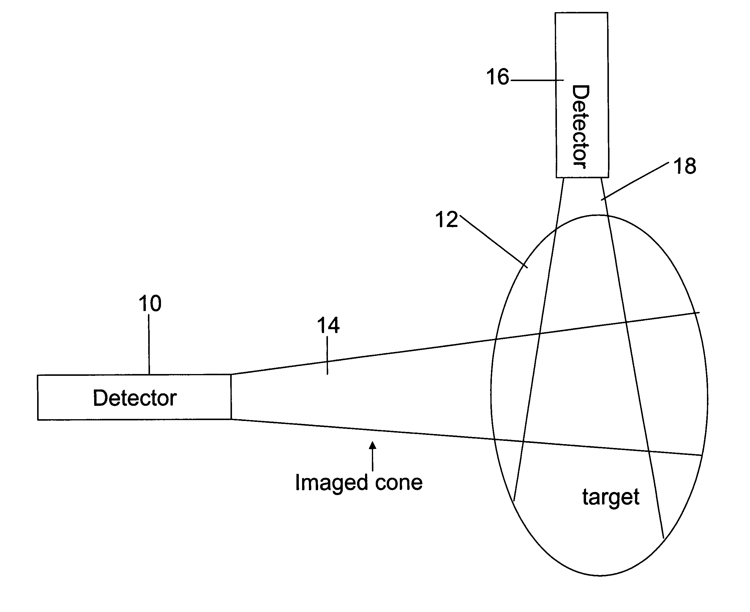
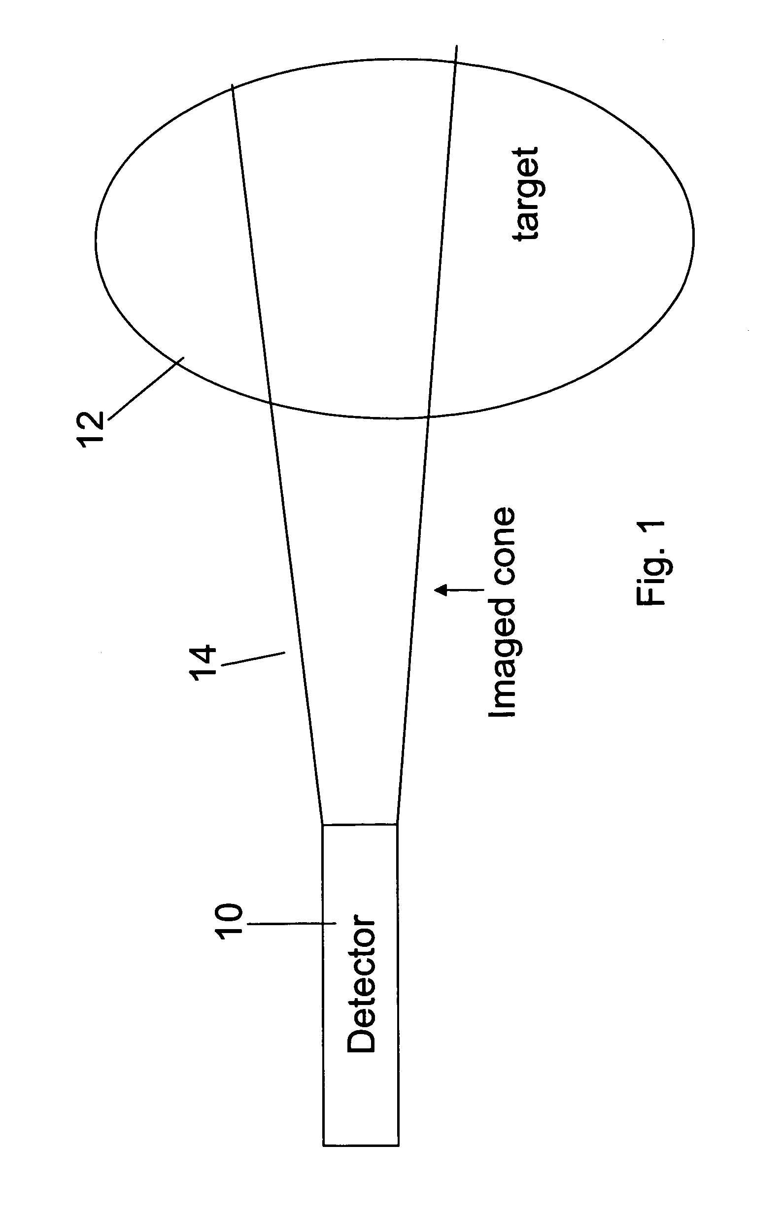
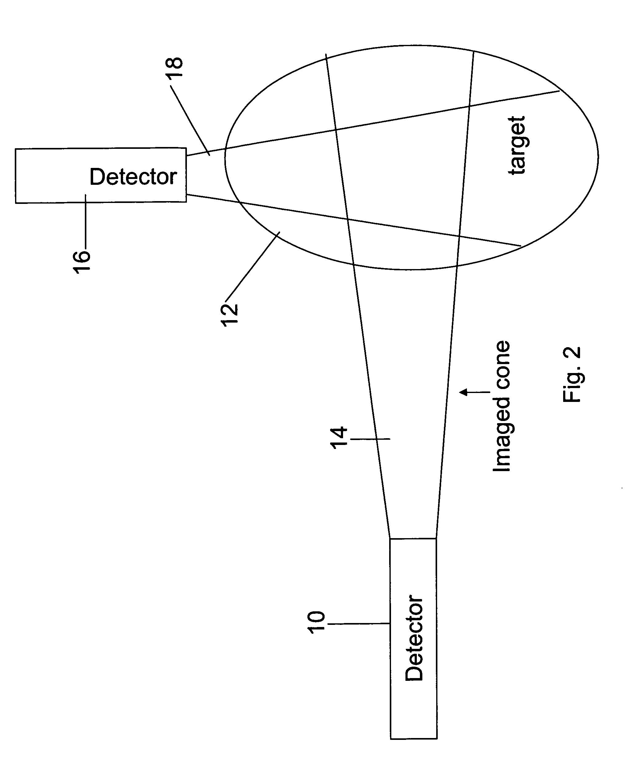
[0067]The present embodiments comprise an apparatus and a method for radiation based imaging of a non-homogenous target area having regions of different material or tissue type or pathology. The imaging uses multi-dimensional data of the target area in order to distinguish the different regions. Typically the multi-dimensional data involves time as one of the dimensions. A radioactive marker has particular time-absorption characteristics which are specific for the different tissues, and the imaging device is programmed to constrain its imaging to a particular characteristic.

[0068]The result is not merely an image which concentrates on the tissue of interest but also, because it is constrained to the tissue of interest, is able to concentrate imaging resources on that tissue and thus produce a higher resolution image than the prior art systems which are completely tissue blind.

[0069]The principles and operation of a radiological imaging system according to the present invention may b...


PUM


Abstract

Apparatus for radiation based imaging of a non-homogenous target area having distinguishable regions therein, comprises: an imaging unit configured to obtain radiation intensity data from a target region in the spatial dimensions and at least one other dimension, and an image four-dimension analysis unit analyzes the intensity data in the spatial dimension and said at least one other dimension in order to map the distinguishable regions. The system typically detects rates of change over time in signals from radiopharmaceuticals and uses the rates of change to identify the tissues. In a preferred embodiment, two or more radiopharmaceuticals are used, the results of one being used as a constraint on the other.

Description

RELATIONSHIP TO EXISTING APPLICATIONS[0001]The present application claims priority from U.S. Provisional Patent Application No. 60 / 535,830 filed Jan. 13, 2004, the contents of which are hereby incorporated by reference.FIELD AND BACKGROUND OF THE INVENTION[0002]The present invention relates to multi-dimensional image reconstruction and, more particularly, but not exclusively to such image reconstruction based on a diffuse radioactive source or sources.[0003]Radiological imaging is generally carried out on a living target, which of course means a mix of tissues in close proximity, if not actually overlapping. The general procedure is to feed the patient with one or more radioactive markers prior to the imaging process. The radioactive markers are taken up by the digestive system and pass into the bloodstream. From the bloodstream the marker passes into the different tissues at varying rates depending on the tissue type. Some tissues absorb markers faster than others and some tissues ...

Claims


Application Information

Patent Timeline
Patent Type & Authority Patents(United States)
IPC IPC(8): G01T1/161G01T1/166
CPCG01T1/161G01T1/166A61B6/481A61B6/4258A61B6/5241A61B19/54A61M5/007A61B6/5217A61B6/4057A61B6/486A61B90/39G16H50/30
Inventor ROUSSO, BENNYNAGLER, MICHAEL
Owner SPECTRUM DYNAMICS MEDICAL LTD
Why Eureka
Try Eureka