Quantitative methods for obtaining tissue characteristics from optical coherence tomography images

a tomography image and optical coherence technology, applied in the field of tissue characterization using optical coherence tomography, can solve the problems that small changes in optical properties that influence the oct signal cannot be readily discerned, and may occur errors in analysis, so as to improve tissue type classification

Inactive Publication Date: 2009-12-10
LIGHTLAB IMAGING
View PDF67 Cites 225 Cited by
  • Summary
  • Abstract
  • Description
  • Claims
  • Application Information

AI Technical Summary

Benefits of technology

[0012]In another aspect the invention relates to a processor-implemented method for identifying tissue components in situ. In one embodiment, the method includes the steps of (a) collecting an OCT dataset of a tissue sample in situ using a probe; (b) measuring an attenuation value and a backscattering value at a point in the tissue sample; and (c) determining a tissue characteristic at a location in the tissue sample corresponding to an image location in an OCT image formed from the OCT dataset in response to the measured attenuation value and backscattering value. The method can include the further step of mapping a pair of coordinates in backscatter-attenuation space to an indicium of the value of the pair of coordinates in the backscatter-attenuation space. The method can include the further step of displaying the indicium corresponding to the measured attenuation and backscatter at the point in the OCT image. The tissue characteristic can be selected from the group consi

Problems solved by technology

Since tissue type is identified by its appearance on the screen, errors may occur in the analysis because certain information (such as ti

Method used


Image

  • Quantitative methods for obtaining tissue characteristics from optical coherence tomography images
  • Quantitative methods for obtaining tissue characteristics from optical coherence tomography images
  • Quantitative methods for obtaining tissue characteristics from optical coherence tomography images

Examples


Embodiment Construction

[0044]The following description refers to the accompanying drawings that illustrate certain embodiments of the invention. Other embodiments are possible and modifications may be made to the embodiments without departing from the spirit and scope of the invention. Therefore, the following detailed description is not meant to limit the invention. Rather, the scope of the invention is defined by the appended claims.

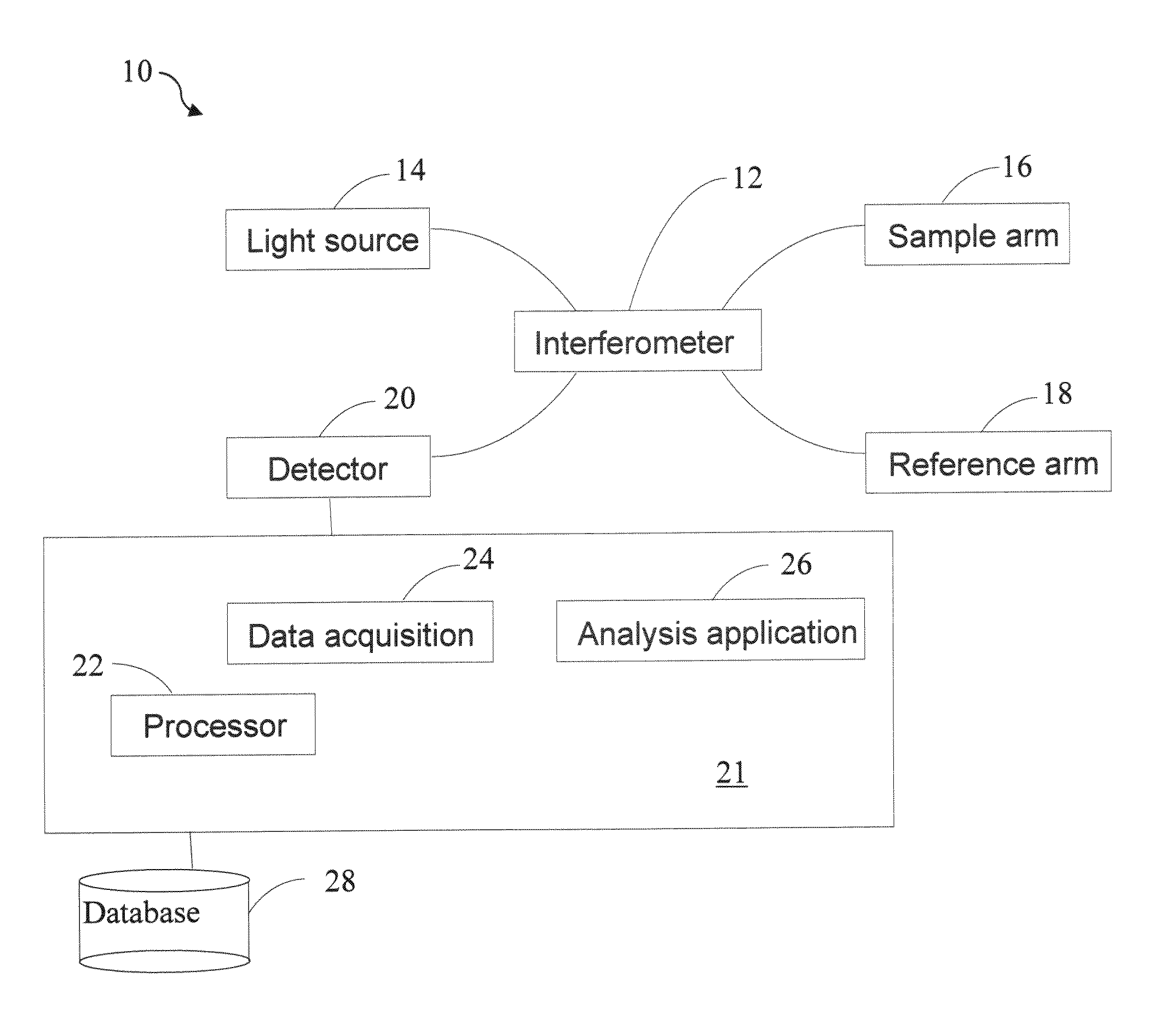
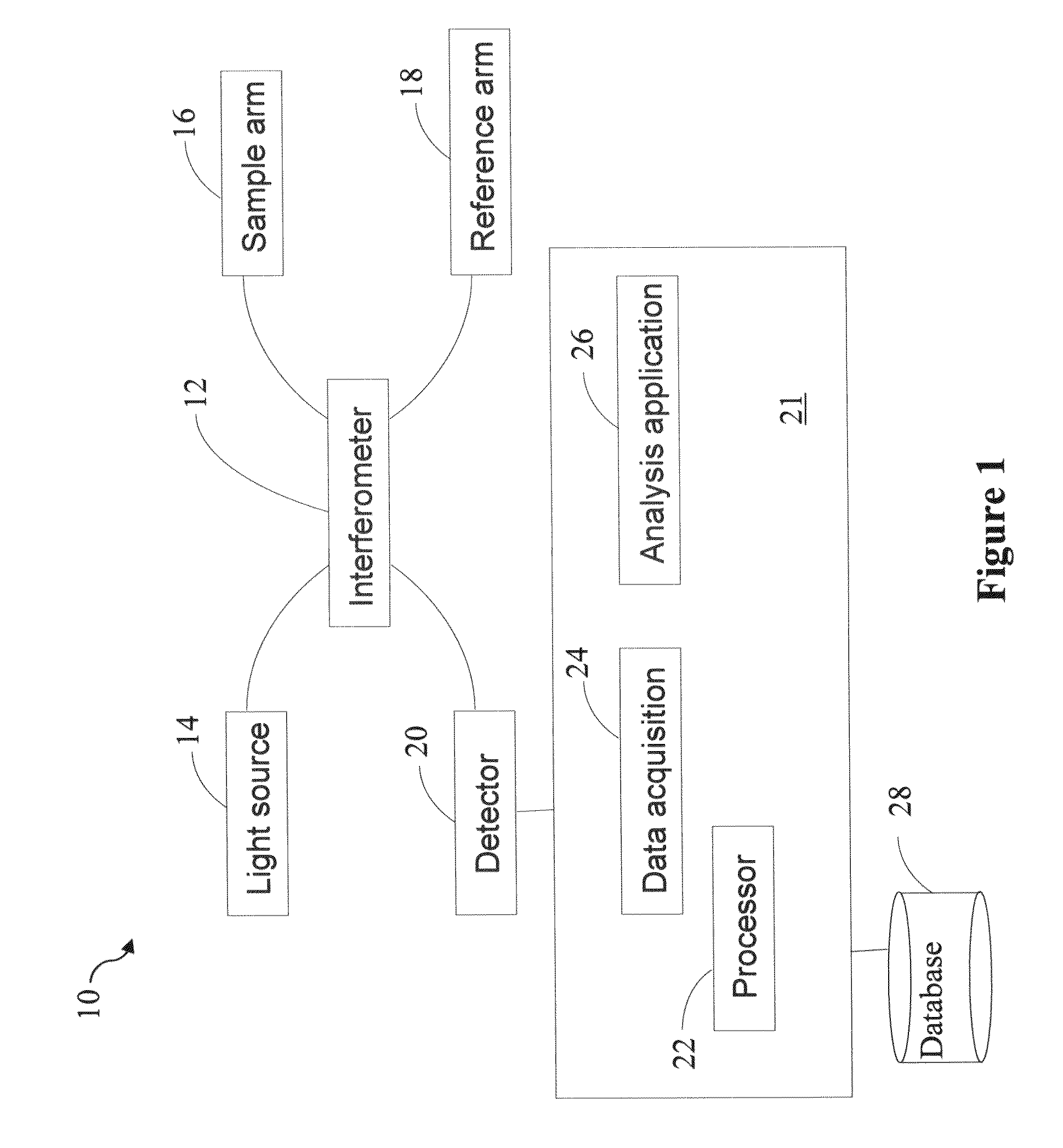
[0045]In general, the invention relates to methods for tissue characterization of vessel walls using optical methods based on what is generally termed low coherence interferometry (LCI), such as, but not limited to optical coherence tomography (OCT) whether in the time or Fourier domain. The methods described herein solve the problems encountered in semi-automatic or automatic tissue characterization application such as optical calibration, artifact removal, generating accurate optical and spatial parameter measurement from regions of interests, tissue segmentations, and sta...


PUM


Abstract

A method and apparatus for determining properties of a tissue or tissues imaged by optical coherence tomography (OCT). In one embodiment the backscatter and attenuation of the OCT optical beam is measured and based on these measurements and indicium such as color is assigned for each portion of the image corresponding to the specific value of the backscatter and attenuation for that portion. The image is then displayed with the indicia and a user can then determine the tissue characteristics. In an alternative embodiment the tissue characteristics is classified automatically by a program given the combination of backscatter and attenuation values.

Description

RELATED APPLICATIONS[0001]This application claims priority to U.S. Provisional Application 61 / 058,077 filed on Jun. 2, 2008, the disclosure of which is herein incorporated by reference in its entirety.FIELD OF INVENTION[0002]This invention provides methods for tissue characterization using optical coherence tomography. Specifically, in part, such characterization can be performed by measuring a tissue's optical and image properties.BACKGROUND[0003]Optical coherence tomography (OCT) is an interferometric imaging technique with widespread applications in ophthalmology, cardiology, gastroenterology and other fields of medicine. The ability to view subsurface structures with high resolution (2-15 μm) through small-diameter fiber-optic probes makes OCT especially useful for minimally invasive imaging of internal tissues and organs. OCT systems can generate images up to 100 frames per second, making it possible to image coronary arteries in the beating heart artery within a few seconds. O...

Claims


Application Information

Patent Timeline
IPC IPC(8): A61B6/00
CPCA61B5/0066A61B5/7264A61B5/6852
Inventor SCHMITT, JOSEPH M.XU, CHENYANG
Owner LIGHTLAB IMAGING
Why Eureka
Try Eureka