
System and method of measuring disease severity of a patient before, during and after treatment

a technology of disease severity and system, applied in the field of diagnosis, prognosis and severity of disease, can solve the problems of reducing diagnostic accuracy, reducing diagnostic accuracy, and reducing the ability to extract objective and quantitatively accurate information from biomedical images, and not keeping pace with the ability to acquire, produce and register images

Active Publication Date: 2005-03-24
SIEMENS MEDICAL SOLUTIONS USA INC
View PDF11 Cites 247 Cited by
  • Summary
  • Abstract
  • Description
  • Claims
  • Application Information

AI Technical Summary

Benefits of technology

[0012] The present invention is provided to solve the above-mentioned problems. According to an aspect of the present invention, there is provided a method of measuring extent and severity of disease in a patient. The method comprises acquiring a first anatomical image data set and a first functional image data set of the patient. The first anatomical image data set and the first functional image data set form a first co-registered composite image data set. At least a volume of interest (VOI) within the first co-registered composite image data set is identified by a user of the system. The first co-registered composite image data set including the VOI is qualitatively and quantitatively analyzed to determine extent and severity of the disease. A second anatomical image data set and a second functional image data set of the patient are acquired. The second anatomical image data set and the second functional image data set form a second co-registered composite image data set. A global, rigid registration is performed on the first anatomical image data set and the second anatomical image data set, such that the first functional image data set and the second functional image data set are also globally registered. At least a VOI within the globally registered image data set using the identified VOI within the first co-registered composite image data set is identified by the user of the system. A local, non-rigid registration is performed on the VOI within the first co-registered composite image data set and the VOI within the globally registered image data set, thereby producing a first co-registered serial image data set. The local, non-rigid registration is more precise than the global, rigid registration. The first co-registered serial image data set including the VOIs is qualitatively and quantitatively analyzed to determine severity of the disease and/or response to treatment of the patient.
[0013] According to another aspect of the pres...

Problems solved by technology

Generally, functional changes often precede structural changes by many days and weeks.
Favorable and unfavorable prognoses are based on predictable changes of structural or functional biomarkers reflecting response of the diseased tissue to treatment.
Since both the pathological and healing processes involve subtle increase or decrease in anatomical morphology, which occur gradually in time, a reliable measure of predictable change may be undetected, thereby reducing diagnostic accuracy.
However, the ability to extract objective and quantitatively accurate information from these biomedical images has not kept pace with the ability to acquire, produce, and register the images.
None of the prior art references disclose the comparison of quantitative functional and structural medical image data to diagnose, prognose or determine the severity of disease using serial examinations obtained before, during, and after treatment of a patient.

Method used


Image

  • System and method of measuring disease severity of a patient before, during and after treatment
  • System and method of measuring disease severity of a patient before, during and after treatment
  • System and method of measuring disease severity of a patient before, during and after treatment

Examples


Embodiment Construction

[0024] In the following detailed description of the preferred embodiment, reference is made to the accompanying drawings which form a part hereof and in which is shown by way of illustrating a specific embodiment in which the invention may be practiced. This embodiment is described in sufficient detail to enable those skilled in the art to practice the invention, and it is to be understood that other embodiments may be utilized and that structural or logical changes may be made without departing from the scope of the present invention. The following detailed description is, therefore, not to be taken in a limiting sense, and the scope of the present invention is defined by the appended claims.

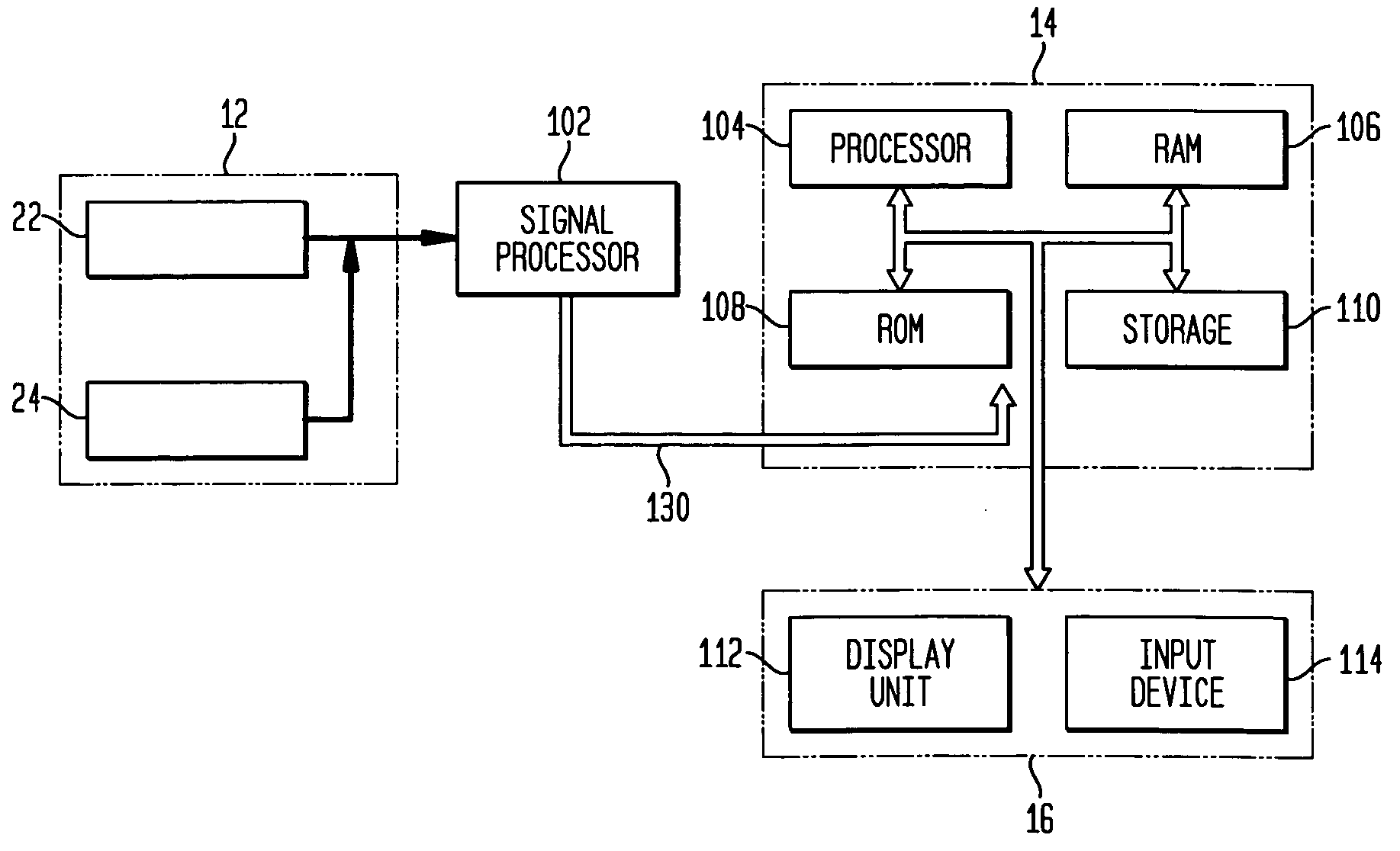
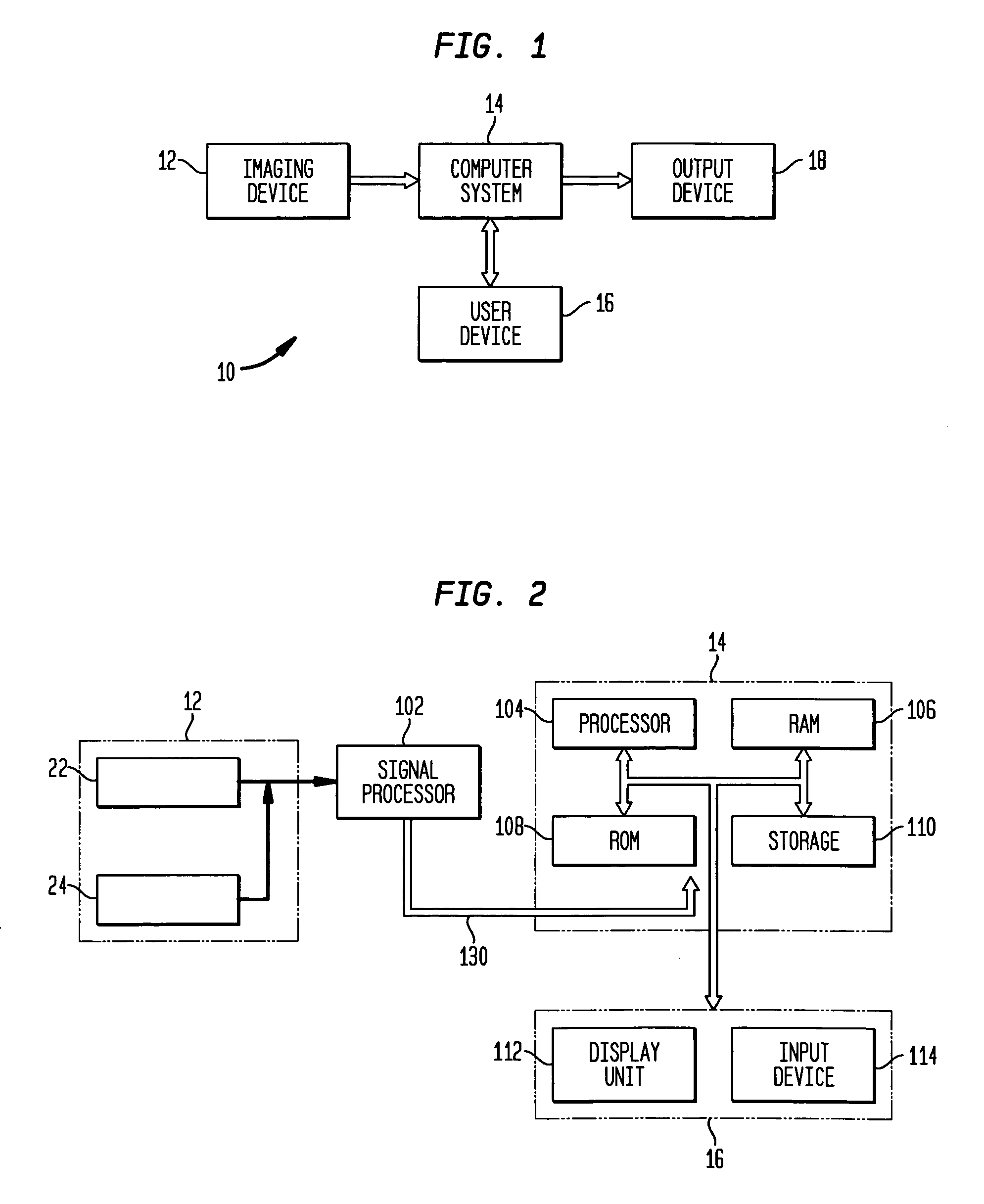
[0025]FIG. 1 schematically shows a system for measuring extent and severity of disease in a patient according to an exemplary embodiment of the present invention. Referring to FIG. 1, a system 10 comprises an imaging device 12 for acquiring anatomical and functional image data sets, a computer...


PUM


Abstract

A system and method of obtaining serial biochemical, anatomical or physiological in vivo measurements of disease from one or more medical images of a patient before, during and after treatment, and measuring extent and severity of the disease is provided. First anatomical and functional image data sets are acquired, and form a first co-registered composite image data set. At least a volume of interest (ROI) within the first co-registered composite image data set is identified. The first co-registered composite image data set including the ROI is qualitatively and quantitatively analyzed to determine extent and severity of the disease. Second anatomical and functional image data sets are acquired, and form a second co-registered composite image data set. A global, rigid registration is performed on the first and second anatomical image data sets, such that the first and second functional image data sets are also globally registered. At least a ROI within the globally registered image data set using the identified ROI within the first co-registered composite image data set is identified. A local, non-rigid registration is performed on the ROI within the first co-registered composite image data set and the ROI within the globally registered image data set, thereby producing a first co-registered serial image data set. The first co-registered serial image data set including the ROIs is qualitatively and quantitatively analyzed to determine severity of the disease and / or response to treatment of the patient.

Description

BACKGROUND OF THE INVENTION [0001] 1. Field of the Invention [0002] The present invention generally relates to the diagnosis, prognosis, and severity of disease using medical images of a patient's body organs or structures of interest. In particular, the present invention relates to a system and method of measuring extent and severity of disease before, during and after treatment. [0003] 2. Description of the Background Art [0004] Disease is any deviation from normal structure or function. A specific disease comprises symptoms manifested as specific biochemical, anatomical or physiological changes. Generally, a patient is non-invasively imaged using various imaging techniques or modalities (e.g., positron emission tomography (PET), single photon emission computed tomography (SPECT), magnetic resonance imaging (MRI), computed tomography (CT), ultrasound, fluoroscopy, x-ray, etc.) in the course of clinical examination to diagnose and determine extent and severity of disease in the pat...

Claims


Application Information

Patent Timeline
IPC IPC(8): A61B5/055A61B6/03G06T7/00
CPCA61B6/032A61B6/037G06T7/0038A61B6/5235G06T7/0012A61B6/469G06T7/38Y10S378/901
Inventor BURCKHARDT, DARRELL DENNIS
Owner SIEMENS MEDICAL SOLUTIONS USA INC
Learn More