Systems, methods and devices for diagnosing sleep apnea

a technology for sleep apnea and systems, applied in the field of systems, methods and devices for diagnosing sleep apnea, can solve the problems of increasing the difficulty of diagnosis, cumbersome equipment, and undiagnosed, and achieve the effect of enhancing the viewing of the airway and enhancing the detection of airway patency

Inactive Publication Date: 2015-02-19
QIU CHUNYUAN
View PDF11 Cites 39 Cited by
  • Summary
  • Abstract
  • Description
  • Claims
  • Application Information

AI Technical Summary

Benefits of technology

[0004]The inventor has recognized disadvantages with the approaches above and herein discloses a wearable hollow nasal tube to be positioned in an upper airway above a constriction point that comprises an end imaging sensor coupled to the nasal tube to collect airway image data; and a controller and communication interface to communicatively link and relay the airway image data during sleep to a select remote device to determine airway obstructions. In one embodiment, the device further comprises an inflatable balloon to anchor the nasal tube such that the end imaging sensor, which is placed at a distal end of the nasal tube, is disposed in an airway image data collection position. Thereby, the device may be positioned within a nasopharnyx above the constriction point with the end imaging sensor angled downward relative to a nose opening, which allows for enhanced viewing of the airway. As described herein, the device may further include one or more biophysical sensors for the enhanced detection of airway patency during OSA screening and diagnosis while an air sampling port configured for monitoring airflow and performing air analysis (e.g., capnography) while visual images of the upper airway are collected may also be included. The device may advantageously further include a head positioning sensor for detecting a device orientation relative to a head position, which allows airway patency to be correlated to sleep positions during sleep studies. Therefore, by placing the nasal tube and end imaging sensor above the point of airway constrictions, breathing patterns can be monitored during sleep without interfering with sleep cycles while the device records and communicates with a remote computing device connected via a network connection. In this way, the device and methods described can be used to advantage for diagnosing OSA by directly imaging the airway, particularly in response to a sleep position, and especially when sleep activities occur remotely.
[0005]As provided herein, in some examples, sleep studies using the disclosed device and systems thereof may be performed in a first location while one or more breathing patterns are remotely analyzed in a second location simultaneously or subsequently, which allows for real-time or early diagnosis of sleep abnormalities in a more convenient manner for both the patient and healthcare professional. For example, a patient may perform the sleep study in the comfort of their own home at night in the U.S. while observations are made and results of the sleep study are analyzed during the day in China (or other locations), or vice versa. Furthermore, in some embodiments, the device may include a clock or timing device, which allows recorded measurements to be time-stamped and thereby synchronized for further data analysis of sleep patterns.

Problems solved by technology

In addition, up to 93% of women and 82% of men having moderate to severe OSA may go undiagnosed for many years if at all due to diagnostic limitations of the currently known methods.
However, PSG involves hooking a patient up to many wires during the sleep period, which may interfere with the sleep cycle and thereby increase the difficulty of diagnosis.
However, such techniques may still require cumbersome equipment to monitor airflows within the upper airway and further lack a means for detecting a sleep position, especially in a remote environment where a trained professional is not present to observe the sleep cycles.

Method used


Image

  • Systems, methods and devices for diagnosing sleep apnea
  • Systems, methods and devices for diagnosing sleep apnea
  • Systems, methods and devices for diagnosing sleep apnea

Examples


first embodiment

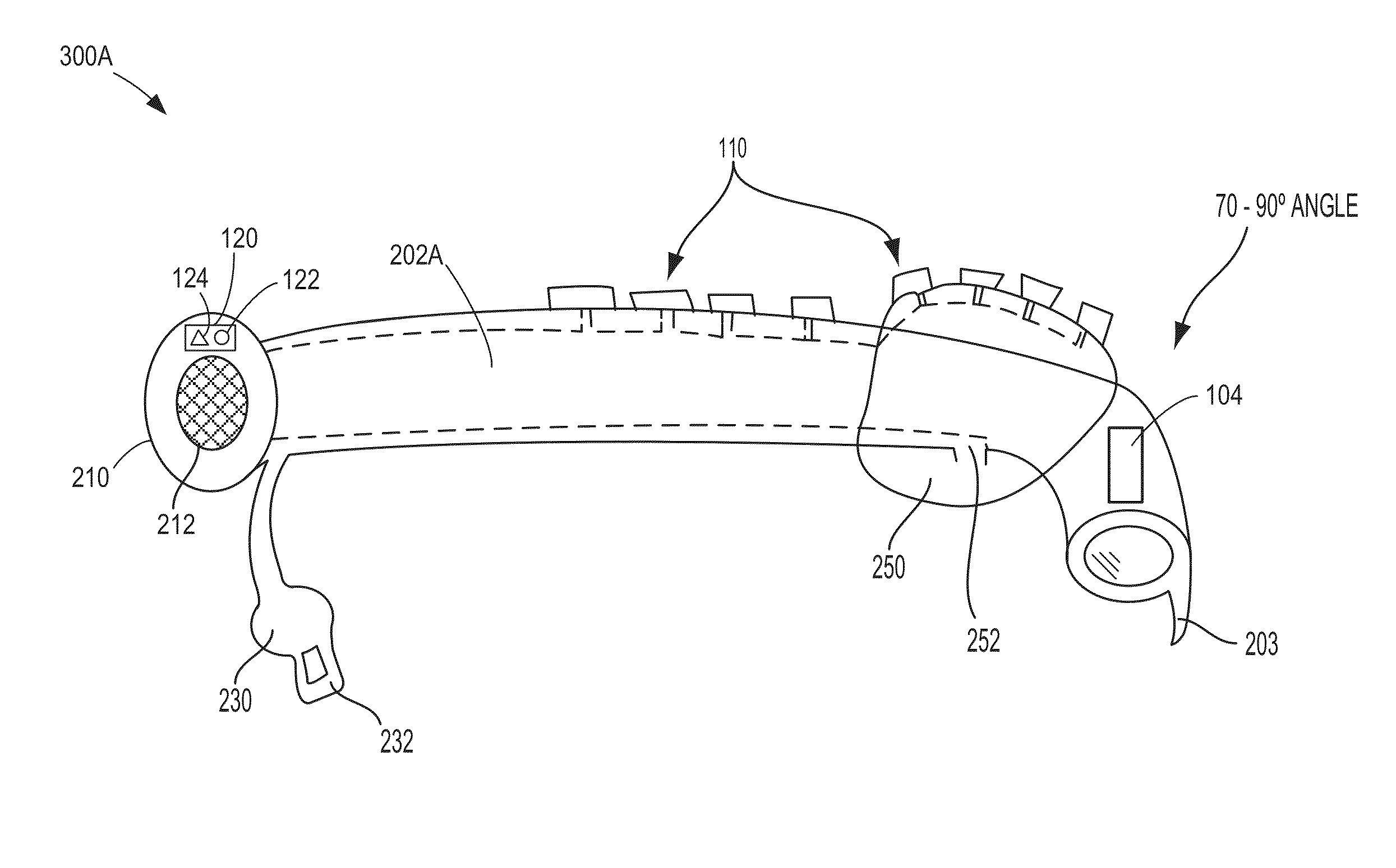
[0032]FIGS. 2A and 2B show example embodiments of OSA diagnostic system 100 in accordance with the present disclosure. For example, FIG. 2A shows airway assessing apparatus 102A with nasal tube 202A inserted into nasal cavity 204. Nasal tube 202A may be a flexible hollow tube adapted for insertion into the nasal cavity. In some examples, nasal tube 202A may include a soft, bendable beak-like tip 203 or other configuration that allows for downward deflection and easier insertion and placement of the device within the cavity above constriction point 206. In the first embodiment shown, airway assessing apparatus 102A includes nose stopper 210 along with mesh 212 that enables air to pass through the hollow device and into the airway. The device may also include pilot balloon 230 with air injection port 232 (or valve) that is used for inflating balloon 250 through inflation opening 252. In the embodiment shown, inflatable balloon 250 may be included along with the nose stopper to anchor ...

second embodiment

[0034]FIG. 2B shows airway assessing apparatus 102B that also includes nasal tube 202B with a soft, bendable end and a flexible beak-like tip 203 for easier insertion and downward deflection to increase the ease of placement within nasal cavity 204 above constriction point 206. In one embodiment, beak-like tip 203 is integrated into the bendable nasal tube and comprises a piece of plastic having the contour of a beak. Thereby, the beak-like tip and nasal tube may form a single, continuous structure. However, this is non-limiting and in other embodiments, the beak-like tip, which is soft and flexible, may also comprise a separate tip configured for addition to the end of the nasal tube and / or may be made of other materials (e.g., rubber) so long as the material is insertable into the body for long periods of time. As noted above, the tip allows for easier and nontraumatic insertion of the tubular airway assessing apparatus into the nasal cavity, since traumatic insertion (and bleedin...


PUM


Abstract

Systems, methods and devices for diagnosing sleep apnea are provided herein. In one example, a device for detecting airway obstructions may be provided where the device may include a wearable hollow nasal tube to be positioned in an upper airway above a constriction point and an end imaging sensor coupled to the nasal tube to collect airway image data. The device further may include a controller and communication interface to communicatively link and relay airway image data to a select remote device to determine airway obstructions. In some examples, a method may be provided including receiving airway image data from a remote end imaging sensor coupled to a nasal tube disposed within an upper airway above a constriction point, and analyzing the airway image data and displaying in a sleep study report the airway image data to identify airway closures related to obstructive sleep apnea.

Description

FIELD[0001]The present description relates generally to systems, methods and devices for diagnosing and monitoring sleep, sleep abnormalities, sleep apnea and related conditions, and more particularly, to systems, methods and devices for observing airway patency during sleep.BACKGROUND AND SUMMARY[0002]Obstructive sleep apnea (OSA) is a disorder in which a person frequently stops breathing during the sleep cycle as the tongue periodically collapses the upper airway to constrict the flow of air therein. Although the prevalence of OSA in the United States is estimated to be 3-7% in men and 2-5% in women, in some populations (e.g., obese patients with a body mass index greater than 28%) the prevalence increases dramatically. In addition, up to 93% of women and 82% of men having moderate to severe OSA may go undiagnosed for many years if at all due to diagnostic limitations of the currently known methods.[0003]Sleep studies designed to detect and diagnose OSA often occur at a sleep stud...

Claims


Application Information

Patent Timeline
Patent Type & Authority Applications(United States)
IPC IPC(8): A61B5/097A61B5/01A61B5/085A61B7/00A61B5/087A61B5/145A61B5/0476A61B5/0402A61B5/083A61B5/00A61B7/04
CPCA61B5/097A61B5/0086A61B5/015A61B5/085A61B5/4818A61B7/003A61B5/742A61B5/14542A61B5/0476A61B5/0402A61B5/0836A61B5/087A61B5/6853A61B7/04A61B5/004A61B5/0826A61B1/233A61B5/0022A61B5/14503A61B5/743A61B7/023A61B5/318A61B5/369
Inventor QIU, CHUNYUAN
Owner QIU CHUNYUAN
Why Eureka
Try Eureka