Anti-complement factor c1q antibodies and uses thereof
a technology of complement factor and anti-complement factor, applied in the field of anti-complement factor c1q antibodies, can solve the problems of neurodegeneration and cognitive declin
- Summary
- Abstract
- Description
- Claims
- Application Information
AI Technical Summary
Problems solved by technology
Method used
Image
Examples
example 1
Production of Anti-C1q Antibodies
[0261]The anti-C1q antibody M1 was generated by Antibody Solutions Inc. (Sunnyvale Calif.) by immunizing C1q knockout mice with human C1q using standard mouse immunization and hybridoma screening technologies (Milstein, C (1999). Bioessays 21: 966-73; Mark Page, Robin Thorpe, The Protein Protocols Handbook 2002, Editors: John M. Walker, pp 1111-1113).
[0262]The anti-C1q antibodies 1C7, 2A1, 3A2, and 5A3 were generated at ImmunoPrecise Ltd (Victoria, BC Canada) by immunizing mice with human C1q protein purified from human plasma (Complement Technology Inc. Tyler Texas, Cat #A-099). In brief, female BALB / c mice were injected intraperitoneal with 25 μg of protein in complete Freund's adjuvant (CFA) on Day 0 and boosts were done with 25 μg of C1q enzyme in incomplete Freund's adjuvant (IFA) on days 21, 42, 52, and a final intravenous boost on Day 63. Four days following the final boost the mice were euthanized, spleens removed and splenocytes were fused w...
example 2
Anti-C1q Antibodies Specifically Bind to C1q
ELISA Screening
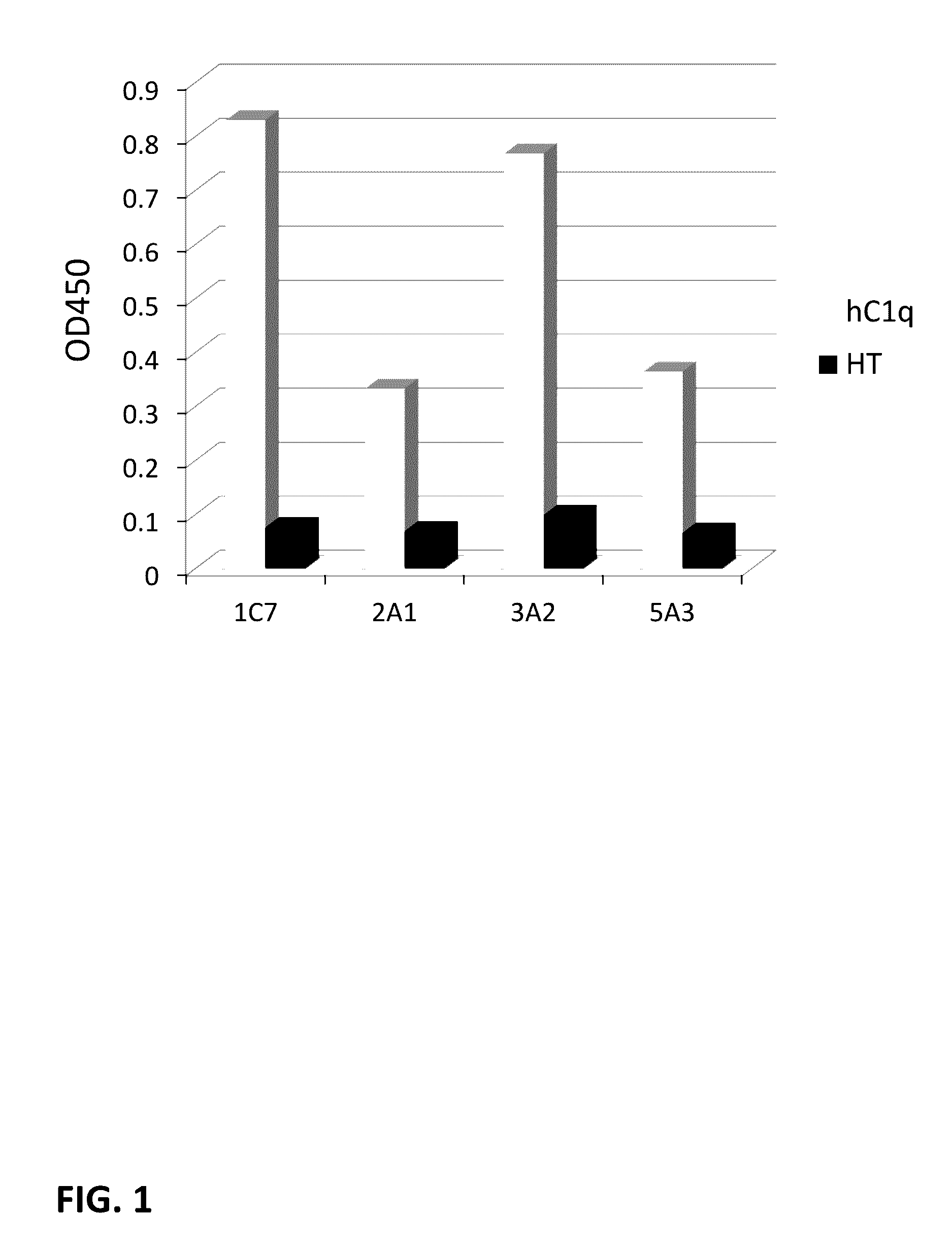
[0264]Anti-C1q antibodies 1C7, 2A1, 3A2, and 5A3 were screened for C1q-binding using standard ELISA protocols.
[0265]Briefly, the day before the assay was performed, 96-well microtiter plates were coated at 0.2 μg / well of C1q-enzyme antigen in 100 μL / well carbonate coating buffer pH 9.6 overnight at 4° C. Control wells were coated with human transferrin. Next, the plates were blocked with 3% milk powder in PBS for 1 hour at room temperature. Then, hybridoma tissue culture supernatants were plated at 100 μL / well for 1 hour at 37° C. with shaking. The secondary antibody (1:10,000 goat anti-mouse IgG / IgM(H+L)-HRP) was applied at 100 μL / well for 1.5 hours at room temperature with shaking. TMB substrate was added at 50 μL / well for 5 minutes at room temperature in the dark. The reaction was stopped with 50 μL / well 1M HCl and absorbance readings were taken at a wavelength of 450 nm.
[0266]Four hybridoma supernatants containing the an...
example 3
Anti-C1q Antibodies Inhibit Complement-Mediated Hemolysis
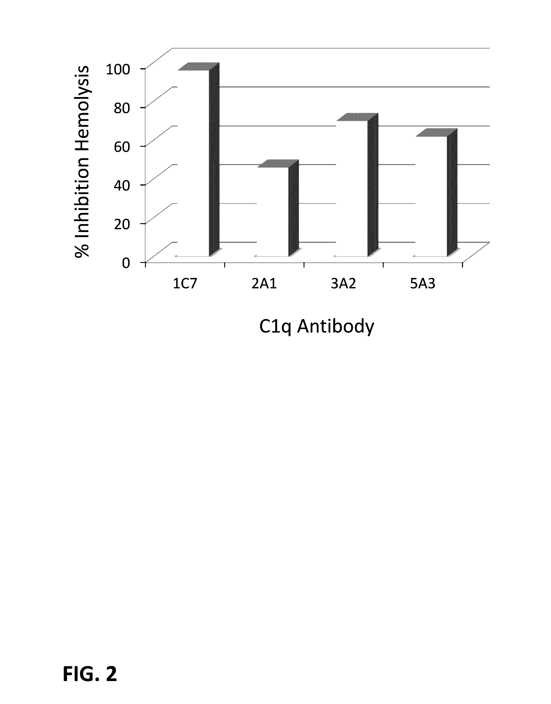
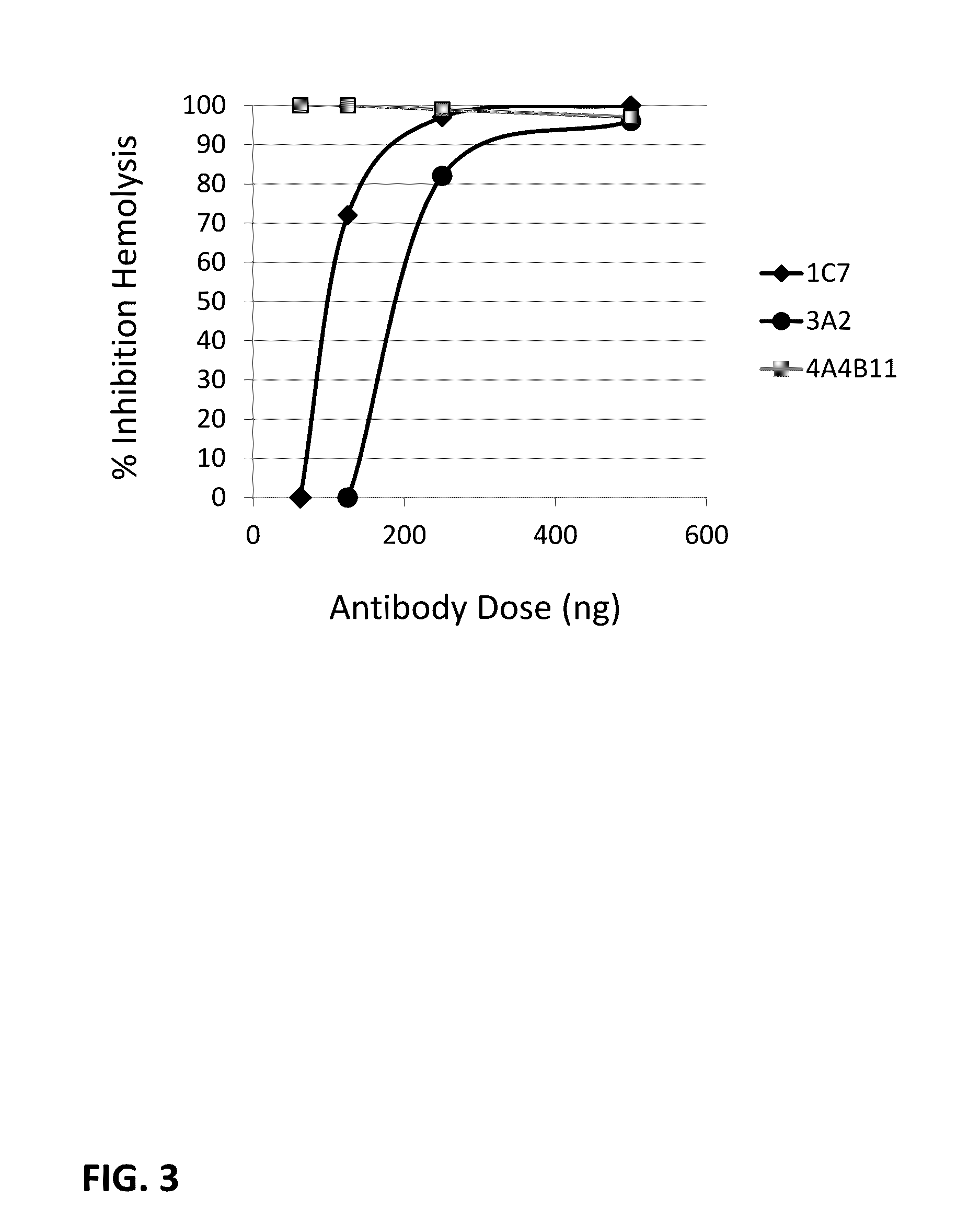
[0274]Anti-C1q antibodies were tested in human and rodent hemolytic assays (CH50) for their ability to neutralize C1q and block its activation of the downstream complement cascade. CH50 assays were conducted essentially as described in Current Protocols in Immunology (1994) Supplement 9 Unit 13.1. In brief, 5 microliters (μl) of human serum (Cedarlane, Burlington, N.C.), 0.625 μl of Wistar rat serum, or 2.5 μl of C57Bl / 6 mouse serum was diluted to 50 μl of GVB buffer (Cedarlane, Burlington, N.C.) and added to 50 μl of the monoclonal antibodies (1 μs) diluted in GVB buffer. The antibody:serum mixture was pre-incubated for 30 minutes on ice and then added to 100 μl of EA cells (2×10⁸ / ml) for rat and human assays, and 4×10⁷ / ml for mouse assays. The EA cells were generated exactly as specified in Current Protocols using Sheeps blood in Alsever's (Cedarlane Cat #CL2581) and hemolysin (Cedalane Cat #CL9000). The EA cells, serum and ...
PUM
| Property | Measurement | Unit |
|---|---|---|
| dissociation constant | aaaaa | aaaaa |
| dissociation constant | aaaaa | aaaaa |
| dissociation constant | aaaaa | aaaaa |
Abstract
Description
Claims
Application Information
 - R&D
- Intellectual Property
- Life Sciences
- Materials
- Tech Scout
- Unparalleled Data Quality
- Higher Quality Content
- 60% Fewer Hallucinations
Browse by: Latest US Patents, China's latest patents, Technical Efficacy Thesaurus, Application Domain, Technology Topic, Popular Technical Reports.
© 2025 PatSnap. All rights reserved.Legal|Privacy policy|Modern Slavery Act Transparency Statement|Sitemap|About US| Contact US: help@patsnap.com


