Method and system for spatial segmentation of anatomical structures

a technology of anatomical structure and spatial segmentation, applied in image analysis, image enhancement, instruments, etc., can solve the problems of large portion of this information going unused and also finding challenges

Active Publication Date: 2017-02-09
PHILIPS MEDICAL SYST TECH
View PDF3 Cites 28 Cited by
  • Summary
  • Abstract
  • Description
  • Claims
  • Application Information

AI Technical Summary

Problems solved by technology

However, in current clinical routines, radiologists estimate the size of anatomical structures on medical images manually, such that a large portion of this information may go unused.
In many cases, such challenges are also found in the segmentation of other soft tissue structures, in particular cysts, nodules, tumors and other lesions.

Method used


Image

  • Method and system for spatial segmentation of anatomical structures
  • Method and system for spatial segmentation of anatomical structures
  • Method and system for spatial segmentation of anatomical structures

Examples


Embodiment Construction

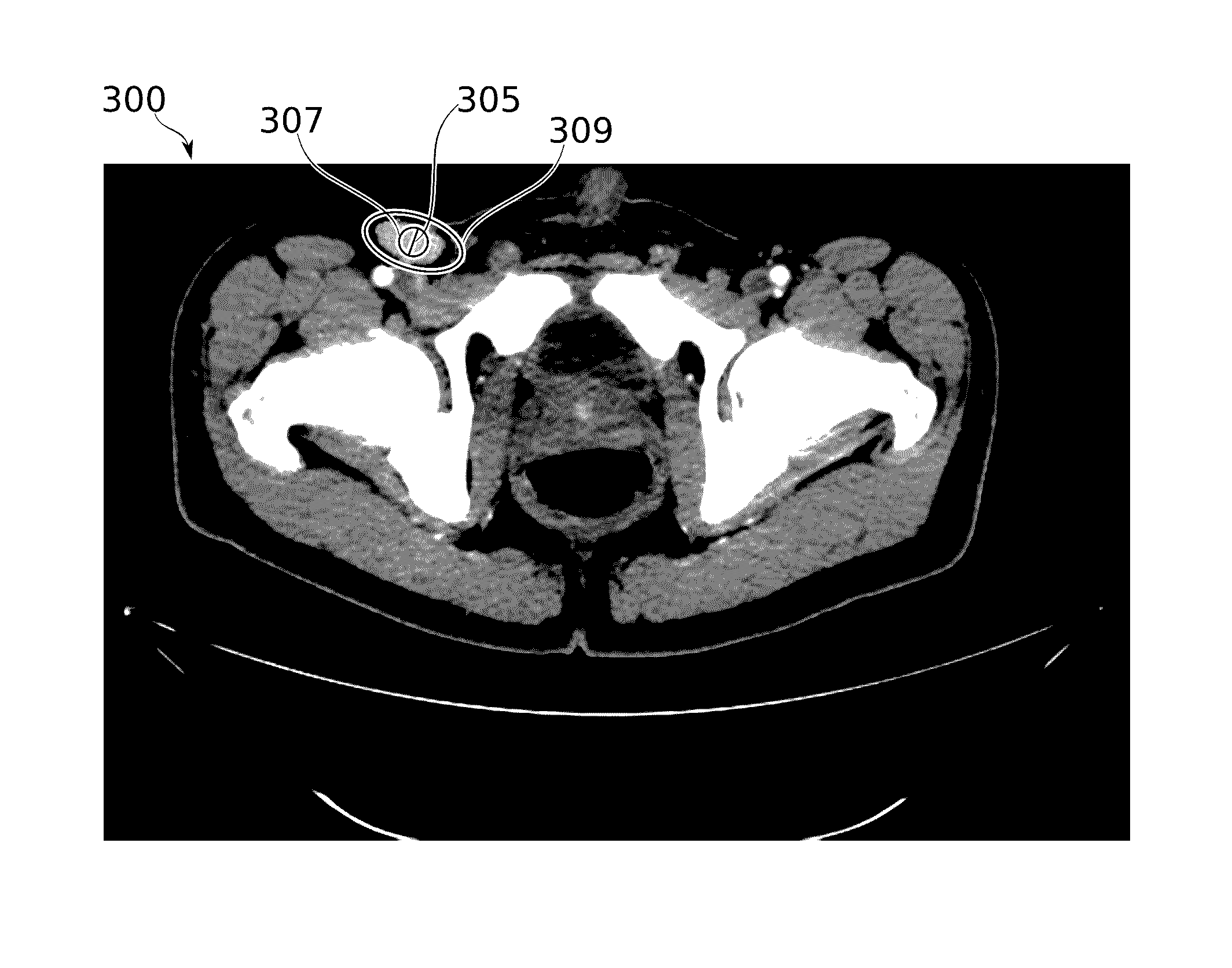
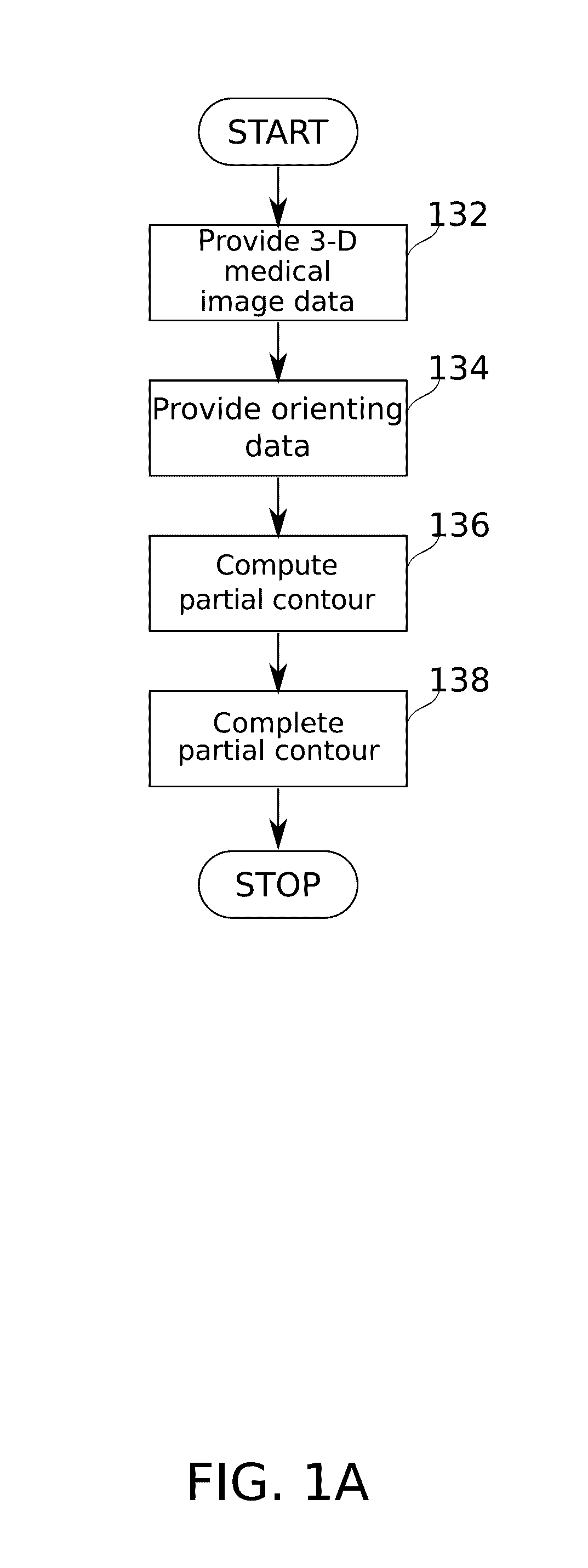
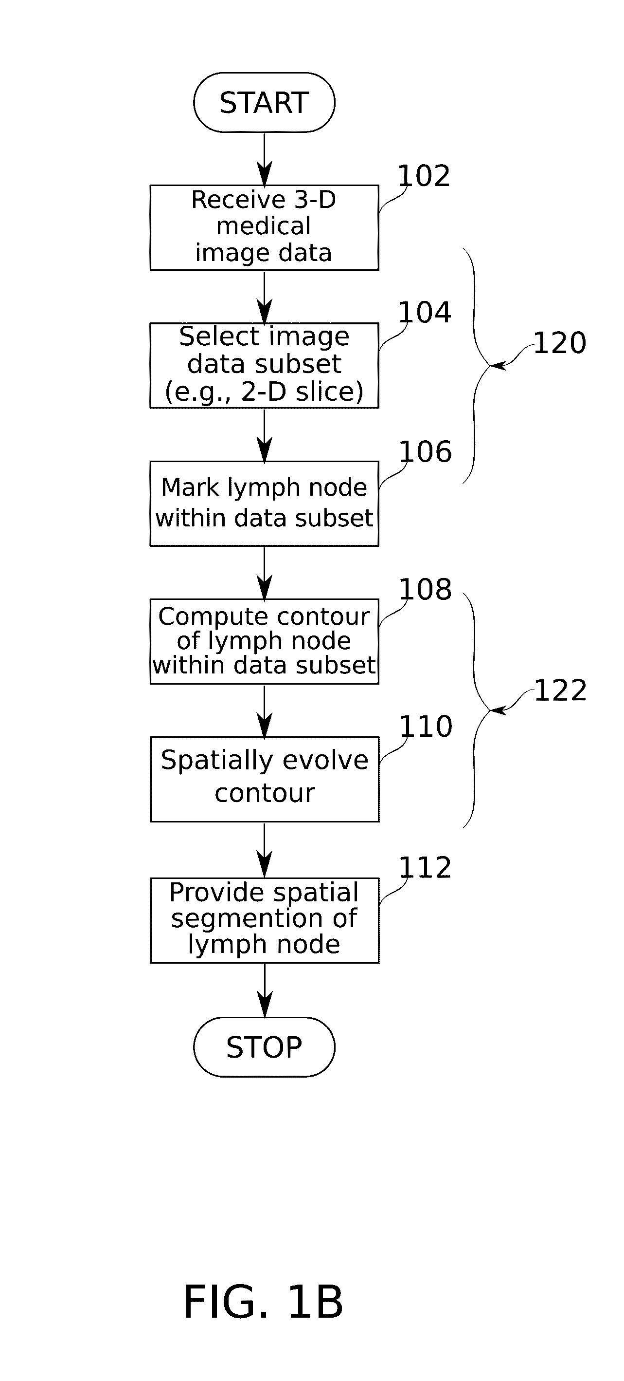
[0090]The present invention, in some embodiments thereof, relates to the field of medical image data segmentation, and more particularly, to semi-automatic spatial segmentation of anatomical structures including, for example: lymph nodes, cysts, tumors, nodules and / or lesions on three-dimensional (3D) medical image data.

[0091]An aspect of some embodiments of the invention relates to determining a spatial segmentation of nodular anatomical structures in a 3-D medical image, based on extrapolation from initial inputs that do not explicitly set the 3-D extents of the nodular structure. For purposes of illustration, many of the descriptions herein relate specifically to lymph nodes. However, it is to be understood that methods of segmentation described herein with respect to lymph nodes are also applicable to other anatomical structures, such as cysts, tumors, nodules and / or other lesions. The methods described herein are potentially of particular utility for segmentation of nodular str...


PUM


Abstract

Spatial segmentation of lymph nodes in a 3-D medical image is automatically determined, based on a set of inputs provided by a user which define a low number of initial conditions for segmentation. In some embodiments, the automation comprises producing a lymph node segmentation from the 3-D image based on a 2-D image slice and a representative line segment on that slice. In some embodiments, segmentation comprises a two tiered approach (2-D segmentation, followed by 3-D segmentation) based on adaptation of the level set framework to the particular conditions of lymph node segmentation.

Description

CROSS REFERENCE TO RELATED APPLICATIONS[0001]This application claims the benefit of U.S. Provisional application U.S. Ser. No. 62 / 201,147 provisionally filed on Aug. 5, 2015, entitled METHOD AND SYSTEM FOR SPATIAL SEGMENTATION OF ANATOMICAL STRUCTURES in the name of Dror et al, which is incorporated herein by reference in entirety.FIELD AND BACKGROUND[0002]The present invention, in some embodiments thereof, relates to the field of medical image data segmentation, and more particularly, to semi-automatic spatial segmentation of anatomical structures including, for example: lymph nodes, cysts, tumors, nodules and / or other lesions on three-dimensional (3D) medical image data.[0003]Lymph nodes, in particular, are bean-shaped organs that play a critical role in the proper functioning of the immune system. They are widely distributed throughout the body, with a primary function to recognize and filter out foreign substances. Lymph nodes become inflamed or enlarged under pathological condi...

Claims


Application Information

Patent Timeline
Patent Type & Authority Applications(United States)
IPC IPC(8): G06T7/00
CPCG06T7/0083G06T7/0012G06T2207/30004G06T2207/10081G06T2207/20156G06T7/12
Inventor DROR, ORANIT D.ENGELHARD, GUY E.
Owner PHILIPS MEDICAL SYST TECH
Why Eureka
Try Eureka