
Method and System for Radiology Structured Report Creation Based on Patient-Specific Image-Derived Information

a structured report and image-derived information technology, applied in the field of automated generation of patient-specific structured report templates, can solve the problems of inability to address the criticism of structured reports by healthcare executives, lack of flexibility of structured reports, and inability to meet the needs of radiologists

Inactive Publication Date: 2017-03-23
SIEMENS HEALTHCARE GMBH
View PDF31 Cites 19 Cited by
  • Summary
  • Abstract
  • Description
  • Claims
  • Application Information

AI Technical Summary

Benefits of technology

The present invention provides a method and system for creating radiology structured reports based on patient-specific information extracted from medical images. The invention automatically generates patient-specific templates for structured reports based on anatomical structures detected in medical images, which allows for more accurate and efficient reporting of healthcare information. Overall, the invention improves the accuracy and efficiency of medical reporting while maintaining the benefits of structured reports.

Problems solved by technology

However, structured reports and template reports have not been widely adopted despite strong evidence that they positively impact the quality of care and communications between radiology departments and referring physicians.
With few notable exceptions (e.g., Mammography), Healthcare executives have been largely unsuccessful in addressing radiologists' criticism of structured reports.
Besides pure and simple resistance to change, the main criticism presented by detractors of structured reports is the lack of flexibility.
More specifically, when presented as templates requiring user selections with mouse clicks from a standardized nomenclature, structured reports have been known to distract radiologists from image interpretation.
In particular, when starting from a templated report instead of a free-form report, radiologists need to first tailor the template to the image currently being interpreted, which typically cannot be done through the usual dictation system.
This may lead to inconvenience in filling the template report for the radiologist who is interpreting the scan.

Method used


Image

  • Method and System for Radiology Structured Report Creation Based on Patient-Specific Image-Derived Information
  • Method and System for Radiology Structured Report Creation Based on Patient-Specific Image-Derived Information
  • Method and System for Radiology Structured Report Creation Based on Patient-Specific Image-Derived Information

Examples


Embodiment Construction

[0015]The present invention relates to methods and systems or creating radiology structured reports based on patient-specific information extracted from medical images. Embodiments of the present invention are described herein to give a visual understanding of the methods for creating radiology structured reports. A digital image is often composed of digital representations of one or more objects (or shapes). The digital representation of an object is often described herein in terms of identifying and manipulating the objects. Such manipulations are virtual manipulations accomplished in the memory or other circuitry / hardware of a computer system. Accordingly, is to be understood that embodiments of the present invention may be performed within a computer system using data stored within the computer system.

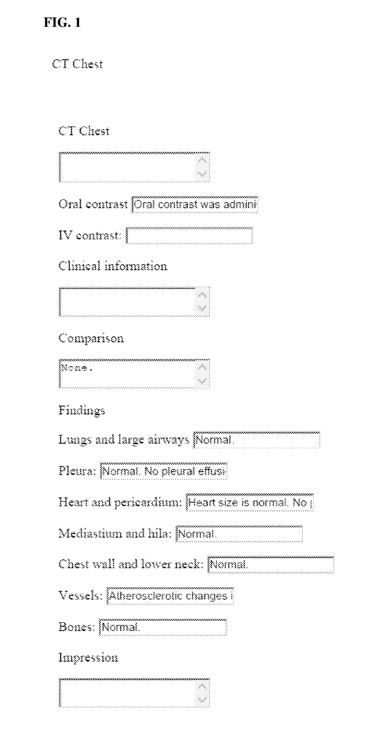
[0016]Radiology structured reports are typically filled out from a template. Examples of some such templates are available on the website: http: / / www.radreport.org, which includes ...


PUM


Abstract

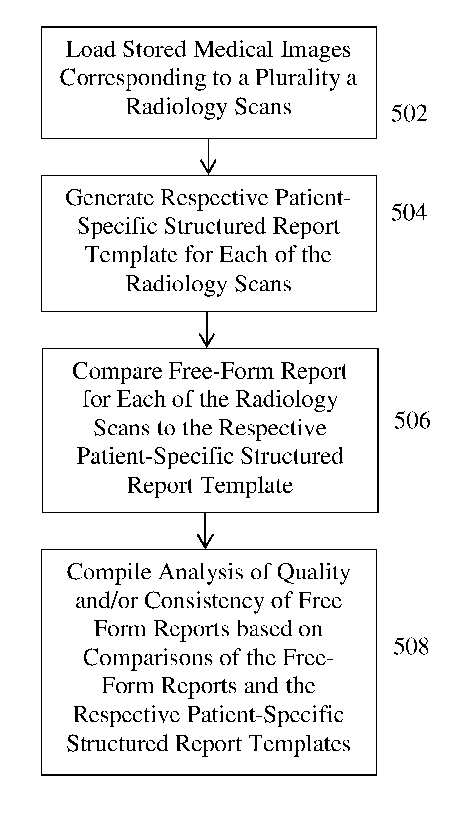
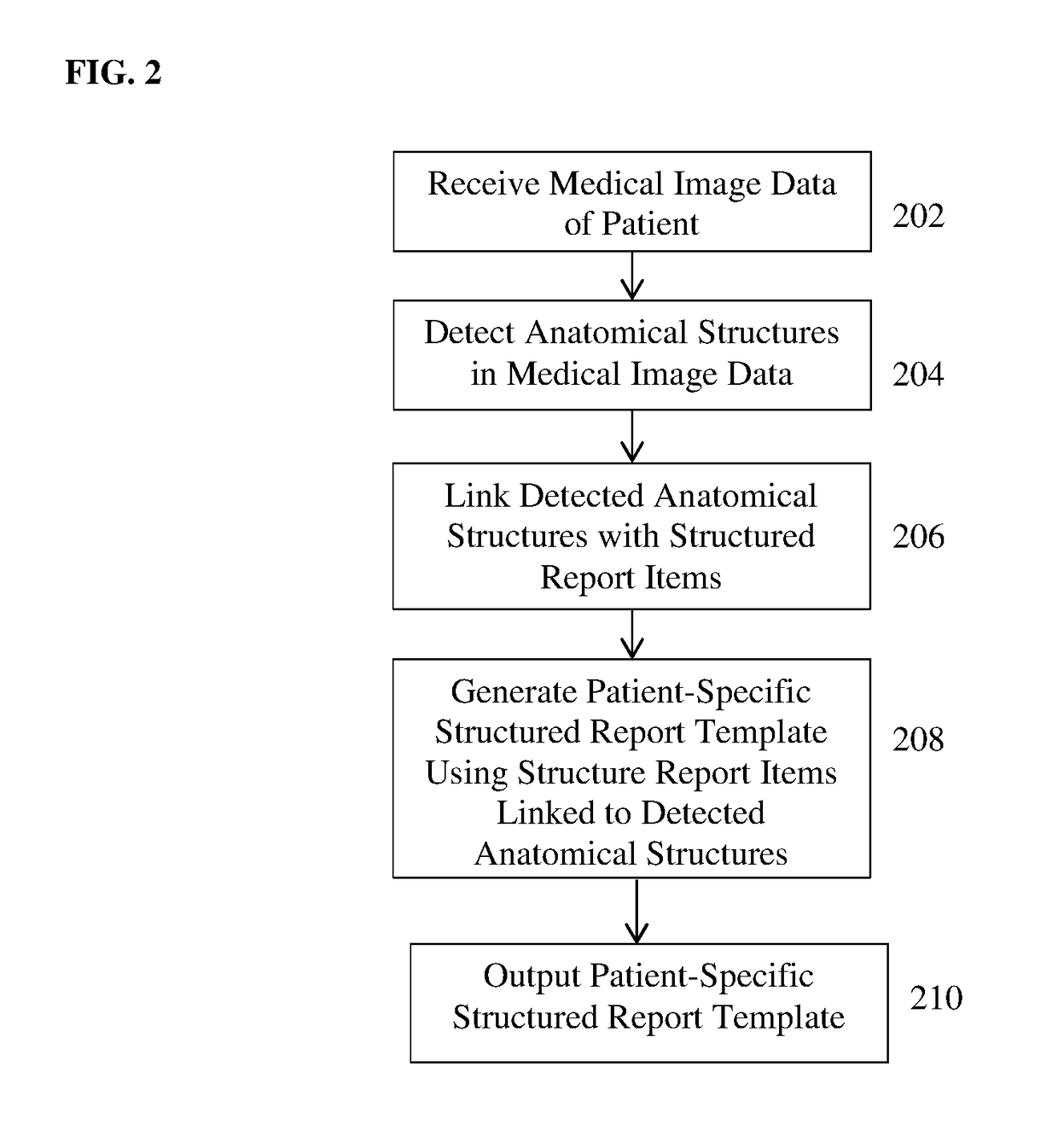
A method and apparatus for automatic generation of a patient-specific structured report template for a radiology scan of a patient is disclosed. Anatomical structures are detected in medical image data acquired in a radiology scan of a patient. The detected anatomical structures are linked to related structured report items in a database of structured report items. A patient-specific structured report template for the radiology scan of the patient is automatically generated using the related structured report items linked to the detected anatomical structures.

Description

[0001]This application claims the benefit of U.S. Provisional Application No. 62 / 222,512, filed Sep. 23, 2015, the disclosure of which is herein incorporated by reference.BACKGROUND OF THE INVENTION[0002]The present invention relates to creation of radiology structured reports, and more particularly, to automatic generation of patient-specific structured report templates based on information extracted from medical images of patients.[0003]Radiology generally refers to the use of medical imaging to diagnose and treat diseases and / or injuries within the body of a patient. Radiology encompasses various medial imaging modalities, such as X-ray, ultrasound, computed tomography (CT), magnetic resonance imaging (MRI), and positron emission tomography (PET). Once medical images of a patient are acquired, a radiologist typically interprets or “reads” the medical images and produces a radiology report, which includes the findings and impressions of the radiologist and / or a diagnosis of the pa...

Claims


Application Information

Patent Timeline
Patent Type & Authority Applications(United States)
IPC IPC(8): G06F19/00
CPCG06F19/321G16H15/00G16H30/20
Inventor FLORIN, CHARLES HENRILIU, DAVID
Owner SIEMENS HEALTHCARE GMBH
Learn More