
Method for dixon mri, multi-contrast imaging and multi-parametric mapping with a single multi-echo gradient-recalled echo acquisition

a multi-echo gradient and echo acquisition technology, applied in the field of magnetic resonance imaging, can solve the problems of many confounding factors, the performance of r2*, swi, lfs and qsm techniques for imaging large volumes might be downgraded, and no process has been developed to fully optimize the acquisition parameters. , to achieve the effect of simple susceptibility

Active Publication Date: 2018-09-13
DRANGOVA MARIA
View PDF0 Cites 5 Cited by
  • Summary
  • Abstract
  • Description
  • Claims
  • Application Information

AI Technical Summary

Benefits of technology

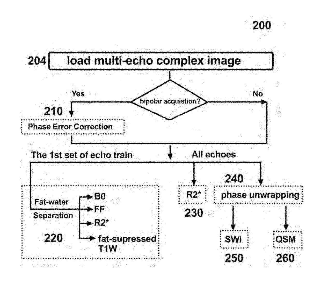
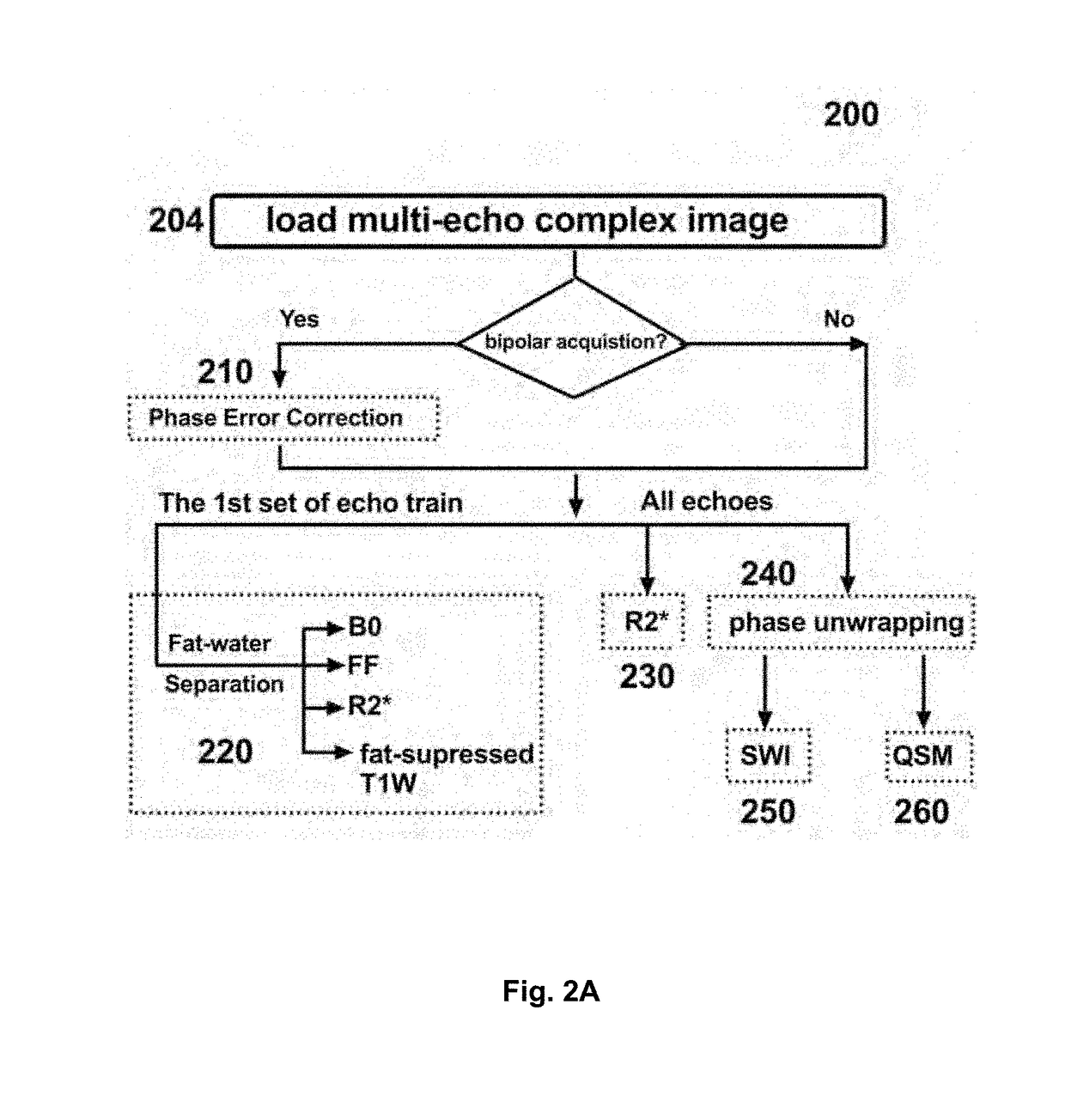
The present invention is a method for magnetic resonance imaging (MRI) that uses a single mGRE sequence with variable echo spacing to capture both water-fat and B0 phase shifts for mapping FF and B0 inhomogeneity. The invention corrects the phase errors associated with bipolar acquisition and uses the derived B0 map to address challenges during phase processing for whole-head QSM and SWI. The invention also presents a fully automated post-processing pipeline that automatically generates co-registered images and maps. The invention is applicable to whole head / neck, liver, knee or even whole body scans with sliding table.

Problems solved by technology

However, the performance of the R2*, SWI, LFS and QSM techniques for imaging large volumes might be downgraded by many confounding factors, particularly, the presence of macroscopic B0 inhomogeneity and fat content (9).
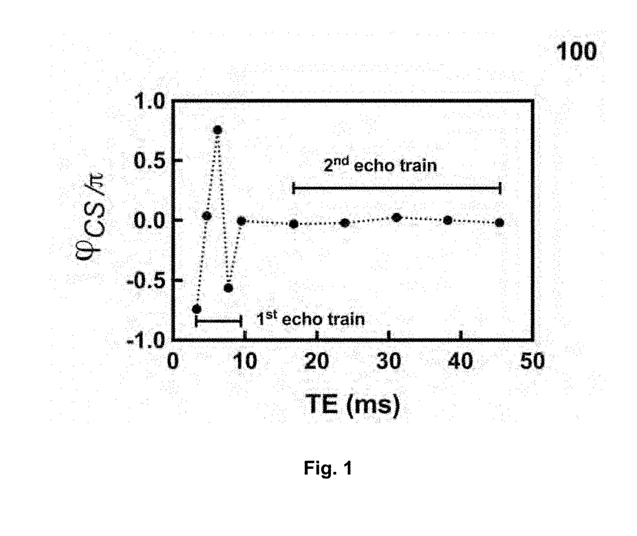
While the concept of acquiring mGRE images with two echo trains has been considered previously (11), no process has been developed which fully optimizes the acquisition parameters, and corrects for the effect of B0 on the phase-sensitive images.

Method used


Image

  • Method for dixon mri, multi-contrast imaging and multi-parametric mapping with a single multi-echo gradient-recalled echo acquisition
  • Method for dixon mri, multi-contrast imaging and multi-parametric mapping with a single multi-echo gradient-recalled echo acquisition
  • Method for dixon mri, multi-contrast imaging and multi-parametric mapping with a single multi-echo gradient-recalled echo acquisition

Examples


examples

[0041]FIGS. 3 to 8 show the intermediate and the final results from Subject #1 as an example of how each step of the pipeline processes the bipolar mGRE data. FIGS. 3 and 4 show the fat-water separation results (Step 220). Without applying phase error correction (i.e., if Step 210 is skipped), the calculated FF maps (FIG. 3a) were not uniform throughout the brain, as expected. The success of Step 210 is demonstrated by the improved quality of the FF images of FIG. 3b, which have a uniform distribution of FF values throughout the brain and no fat-water swaps are present in the brain or neck regions, while flow artifacts are observable as suggested by arrows.

[0042]FIG. 4 shows a few examples when using the afore-determined FF map to generate water-only (FIG. 4b) and fat-only (FIG. 4c) images from the anatomical images (FIG. 4a). These results suggest that uniform fat signal suppression of the anatomical images is obtained; slices covering the region around the optic nerve were selecte...


PUM


Abstract

To perform Dixon MRI, generate multi-contrast images, and extract multi-parametric maps, this invention presents a multi-echo gradient echo protocol with two sets of echo trains. An example implementation of the invention at 3 T acquires a short-TE train (ΔTE˜1.2 ms, TE <10 ms), which is used to map B0 inhomogeneity and proton density fat fraction (FF), and a second—susceptibility sensitive—long-TE train (16 ms<TE<45 ms) will enable quantification of local frequency shift (LFS) and susceptibility. The presented pipeline automatically generates co-registered images and maps with / without fat-suppressed, including magnitude- and complex-based FF map, B0 map, anatomical images, brain mask, R2* map, unwrapped phase maps for each echo, susceptibility-sensitive images (SWI, LFS and quantitative susceptibility) for each echo, mean susceptibility-sensitive images for each echo-train. The invention is directly applicable to whole head / neck, liver, knee or even whole body scans with sliding table.

Description

CROSS-REFERENCE TO RELATED APPLICATION[0001]This application claims priority of U.S. Provisional application Ser. No. 62 / 470,164, filed Mar. 10, 2017. The content of the prior application is incorporated herein by reference in its entirety.TECHNICAL FIELD[0002]This disclosure relates to magnetic resonance imaging (MR), particularly, generating Dixon MRI, multi-contrast imaging and multi-parametric mapping by processing the signals collected with a single multi-echo gradient echo acquisition.BACKGROUND[0003]Multi-echo gradient echo (mGRE) sequences have been widely adapted in clinical and scientific practice for different purposes due to their capability of generating multi-contrast images and extracting multi-parametric maps. Using the magnitude mGRE data, R2* relaxivity (R2*=1 / T2*) mapping techniques (1) have been used to quantify blood oxygenation level dependent functional MRI, detect and track of super-magnetic iron oxides, visualize abnormalities of the articular knee, assess i...

Claims


Application Information

Patent Timeline
Patent Type & Authority Applications(United States)
IPC IPC(8): G01R33/56G01R33/561G01R33/24
CPCG01R33/5607G01R33/5616G01R33/243G01R33/5608G01R33/4828G01R33/50G01R33/56545G01R33/56554
Inventor LIU, JUNMINDRANGOVA, MARIA
Owner DRANGOVA MARIA
Learn More