Realistic electro-anatomical model of the mammalian his/purkinje system

an electro-anatomical model and mammalian technology, applied in educational models, medical science, surgery, etc., can solve the problems of increased risk of heart failure, increased risk of atrial fibrillation and overall mortality, and difficulty in achieving his pacing

Active Publication Date: 2019-06-06
ST JUDE MEDICAL CARDILOGY DIV INC
View PDF0 Cites 2 Cited by
  • Summary
  • Abstract
  • Description
  • Claims
  • Application Information

AI Technical Summary

Problems solved by technology

While effective, this technique can cause abnormal electrical activation sequences resulting in mechanical ventricular dyssynchrony and an increased risk of heart failure, atrial fibrillation and overall mortality.
While the improved results obtainable with His pacing have been recognized, in practice His pacing is difficult to achieve because the bundle of His is very small and difficult to locate and access with available pacing tools.
As a result of its small size and weak electrical signal, the bundle of His is extremely difficult to find with a conventional pacing lead.
Moreover, once the bundle of His has been located, it is difficult to maintain the position of the lead while it is being affixed to the cardiac tissue.
The difficulties involved in locating the bundle of His and affixing a pacing lead thereto result in lengthy implantation procedures.
Both attempts to develop tools for His pacing, and the training of users in techniques for implanting His pacing leads, have been limited by the inadequacy of development models.
Models with approximately appropriate anatomy and physiology have been limited to large mammals such as pigs or dogs.
The use of these models is hindered by inconvenience, both in terms of expense and the inability to see directly where the lead is going, and by significant anatomic and physiologic differences between the best available animal models and humans.

Method used


Image

  • Realistic electro-anatomical model of the mammalian his/purkinje system
  • Realistic electro-anatomical model of the mammalian his/purkinje system
  • Realistic electro-anatomical model of the mammalian his/purkinje system

Examples


Embodiment Construction

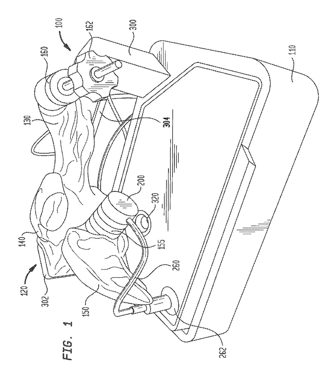
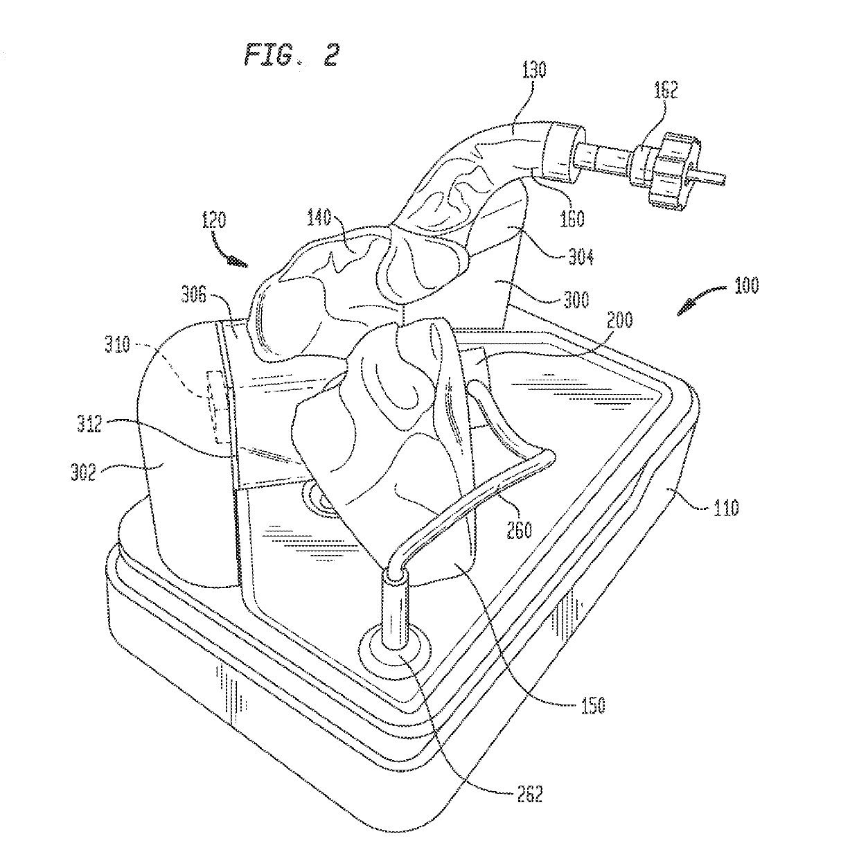
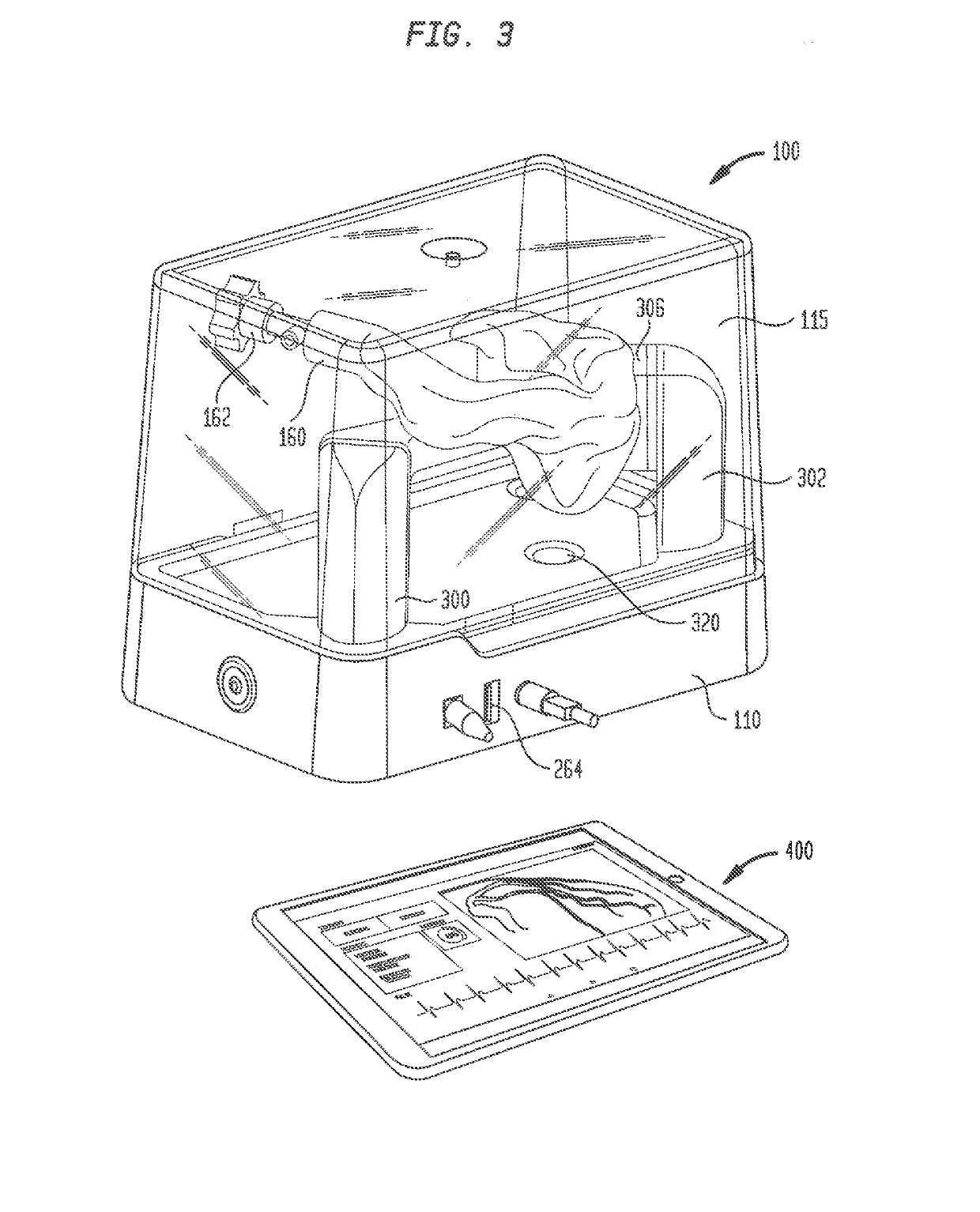
[0026]An electro-anatomical model 100 according to the present disclosure is shown in FIGS. 1-3. Model 100 includes three main components, namely, an anatomically accurate shell 120, an electrically active plug insert 200, and a circuit for producing a simulated cardiac signal, such as the His signal.

[0027]Shell 120 is mounted on a base 110 that houses a circuit board for generating desired electrical signals, as well as other electronics for operating model 100. A removable cover 115 may mate with base 110 to cover shell 120 and protect same during shipment of model 100 and periods of nonuse. Shell 120 need only be as accurate as is needed for training or testing purposes. Thus, it need not look like, be oriented like, or be sized like the native anatomy so long as it is sufficiently anatomically accurate to permit its effective use. In one specific embodiment, as illustrated herein, shell 120 generally reproduces the anatomy of the right side of the human heart. Thus, referring to...


PUM


Abstract

An electro-anatomical model of the mammalian His / Purkinje system includes a shell simulating the anatomy of at least a portion of a mammalian heart. The shell has a hollow interior and a first port providing an aperture to the interior of the shell. A plug is inserted in the first port so that a surface of the plug is exposed to the interior of the shell. An electrical circuit provides signals to electrodes in the plug which simulate the electrical signals generated by the bundle of His / Purkinje system in vivo. A second port provides access to the interior of the shell for an introducer catheter to locate the simulated bundle of His and to insert a pacing lead therein. The model is useful for developing tools for His pacing and for training users in techniques for implanting His pacing leads.

Description

CROSS-REFERENCE TO RELATED APPLICATIONS[0001]The present application claims the benefit of the filing dates of United States Provisional Patent Application Nos. 62 / 594,130, filed Dec. 4, 2017, and 62 / 598,518, filed Dec. 14, 2017, the disclosures of which are hereby incorporated by reference herein.FIELD OF THE DISCLOSURE[0002]The present disclosure relates to cardiac resynchronization therapy (CRT), and more particularly to pacing of the His bundle in the heart of a patient. Still more particularly, the present invention relates to an anatomical model for facilitating the development of tools for use in His pacing, as well as for training users in techniques for implanting His pacing leads in patients.BACKGROUND OF THE DISCLOSURE[0003]Cardiac rhythm management systems are useful for electrically stimulating a patient's heart to treat various cardiac arrhythmias. The current standard of care is to pace the right ventricle by myocardial stimulation. In this technique, pacemaker leads ...

Claims


Application Information

Patent Timeline
Patent Type & Authority Applications(United States)
IPC IPC(8): G09B23/30A61B34/10A61N1/365
CPCG09B23/30A61B34/10A61N1/365A61B2018/00351A61B2017/00053A61B2034/105A61N1/056A61B2017/00707
Inventor KRAMER, SCOTT A.BELK, PAUL A.TANTALEAN, II, JUAN CARLOSSEEKER, GAVIN P.SENARITH, PATRICK P.STOERMER, ERICH W.BROMAN, DAVID J.
Owner ST JUDE MEDICAL CARDILOGY DIV INC
Why Eureka
Try Eureka