Distal Biceps Tendon Repair Device

a distal biceps and tendons technology, applied in the field ofmusculoskeletal repair, can solve the problems of increasing the risk of heteropic ossification, damage to the supinator muscle, and not having a device currently available to allow for perfect anatomical repair of distal biceps tendons, and reducing muscle strength after surgery

Active Publication Date: 2019-06-27
UNIVERSITY OF ROCHESTER
View PDF0 Cites 0 Cited by
  • Summary
  • Abstract
  • Description
  • Claims
  • Application Information

AI Technical Summary

Problems solved by technology

Therefore, while the two incision posterior approach allows anatomically correct repair and provides a superior mechanical attachment technique to that of the one incision anterior approach, it involves the downside of two incisions, increased risk of heteropic ossification and damage to the supinator muscle; a procedure that many surgeons are reluctant to undertake.
Therefore, anterior single incision surgeries are much more commonplace, there is not a device currently available to allow for perfect anatomical repair of distal biceps tendons without the downside of the PA (two incision) approach.
Both the non-anatomic repair that is typically performed with the one incision anterior approach as well as the anatomic repair that is typically performed with the two incision posterior approach result in decrease in muscle strength after surgery: the former approach due to non-anatomic reattachment of the tendon and the latter approach due to cutting of the supinator muscle.
Due to increased risk of heterotopic ossification and damage to supinator muscle, anatomic repair using a two incision posterior approach is still a risky procedure, and one that many surgeons shy away from in favor of the non-anatomic single incision anterior approach.

Method used


Image

  • Distal Biceps Tendon Repair Device
  • Distal Biceps Tendon Repair Device
  • Distal Biceps Tendon Repair Device

Examples


Embodiment Construction

[0025]For a general understanding of the present invention, reference is made to the drawings. In the drawings, like reference numerals have been used throughout to designate identical elements.

[0026]The present invention will be described by way of example, and not limitation. Modifications, improvements and additions to the invention described herein may be determined after reading this specification and viewing the accompanying drawings; such modifications, improvements, and auditions being considered included in the spirit and broad scope of the present invention and its various embodiments described or envisioned herein.

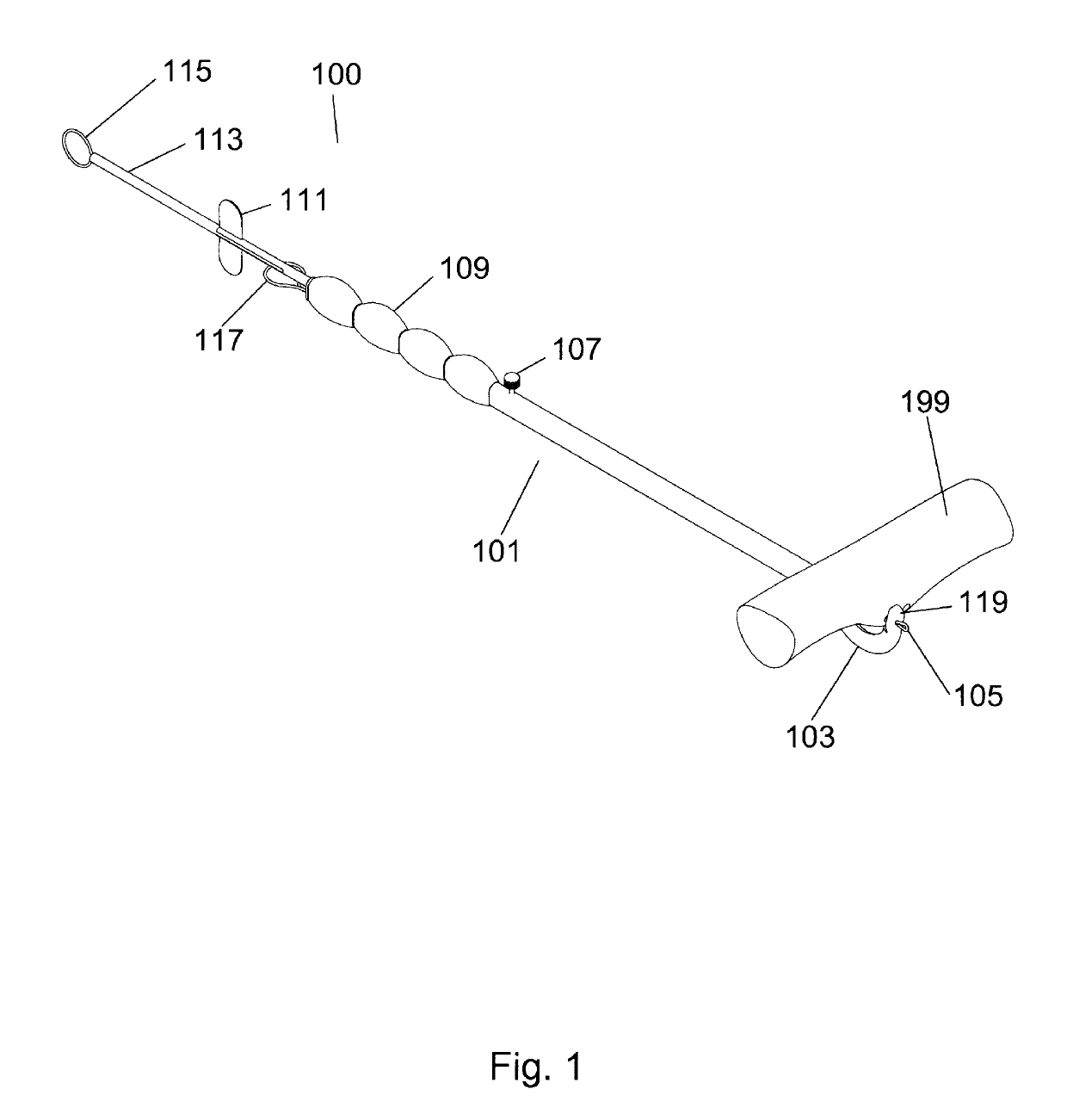
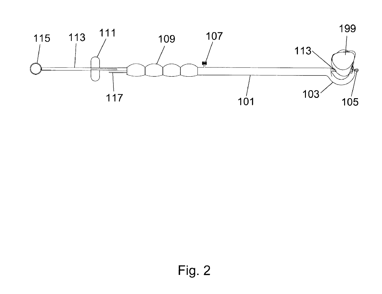
[0027]The distal biceps tendon repair device allows for the repair and anatomically correct reattachment, repair and healing of a distal biceps tendon tear using a single incision anterior surgical approach. Such an approach provides for superior supination strength post-surgery due to the tendon reattachment point used, while reducing the risk associated with a...


PUM


Abstract

A distal biceps tendon repair device is disclosed. The device allows for anatomically correct biceps tendon repair using a single incision anterior approach without the risk associated with a double incision posterior approach. The distal biceps tendon repair device eliminates the necessity of cutting supinator muscle and greatly reduces the risk of contact or damage to vital structures such as arteries or nerves. The device has a bone drill guide mechanically coupled to a cannulated bone hook. A retrieval wire passes through the cannulated bone hook to grasp a suture or suture guide that has passed through a cannulated drill bit retained by the bone drill guide, the retrieval wore brings the suture through the cannulated hone hook, thus bringing the suture through and around the hone for subsequent anatomically correct attachment and placement of sutures used to reattach the distal biceps tendon to the bone.

Description

CROSS REFERENCE TO RELATED PATENT APPLICATIONS[0001]This application claims priority to U.S. Patent Application Ser. No. 62 / 375,354 filed Aug. 15, 2016 entitled “Distal Biceps Tendon Repair Device” by Dr. Ilya Voloshin, M.D., and to International Application Number PCT / US17 / 46828 filed Aug. 14, 2017, the entire disclosures of which are incorporated herein by reference in their entirety.BACKGROUND OF THE INVENTION1. Field of the Invention[0002]This invention relates generally to musculoskeletal repair, and more specifically to a device that assists a surgeon in distal biceps tendon repair surgery using an anterior incision that provides optimal tendon reattachment with minimal loss of supination strength and less risk to the posterior interosseous nerve.2. Description of Related Art[0003]To regain use distal biceps tendon tears need to be repaired in patients who are not sedentary. There are currently two main surgical approaches to distal biceps tendon repair. There is a one incisio...

Claims


Application Information

Patent Timeline
Patent Type & Authority Applications(United States)
IPC IPC(8): A61B17/17A61F2/08
CPCA61B17/1714A61F2/0805A61B17/1739A61B2017/0464
Inventor VOLOSHIN, ILYA
Owner UNIVERSITY OF ROCHESTER
Why Eureka
Try Eureka