Methods for Preparing Therapeutically Active Cells Using Microfluidics

a technology of microfluidics and cells, applied in the field of cells, can solve the problems of labor-intensive process for personalized therapy preparation of cells, and achieve the effects of less senescence, stable cells, and convenient size-based microfluidic separation

Active Publication Date: 2020-02-20
UNIV OF MARYLAND BALTIMORE +2
View PDF0 Cites 9 Cited by
  • Summary
  • Abstract
  • Description
  • Claims
  • Application Information

AI Technical Summary

Benefits of technology

[0012]In a preferred embodiment, the carriers have on their surface an affinity agent (e.g., an antibody, activator, hapten, aptamer, nucleic acid sequence, or other compound) that allows the carriers to bind directly to the target cells with specificity. Alternatively, there may be an intermediary protein, cell, or other agent that binds to both the target cell and carrier with specificity. For example, antibodies may be used that recognize surface antigens on target cells and that also bind with specificity to carriers (e.g., due to that presence of a second antibody on the carrier surface, avidin / biotin binding or some other similar interaction). In addition, target cells may sometimes interact with specificity with other cells to form a complex and in so doing, the other cells may serve as a biological carrier, i.e., they may increase the effective size of the target cell and thereby facilitate its separation from uncomplexed cells. For example, human T cells may interact with sheep erythrocytes or autologous human erythrocytes to form a rosette of cells that can then be purified as a complex. Alternatively, other carriers may bind with specificity to cells in such a rosette to further promote a size based separation.
[0093]Compared to a procedure in which cells are isolated or concentrated by centrifugation, CAR T cells obtained by microfluidic separation should be available for use by a patient at least one day (and preferably, at least 3, 5 or 10 days) earlier. Overall, the time necessary to produce a sufficient number of CAR T cells for treatment should be at least 10% (and preferably at least 20% or 30%) shorter than when the same method is carried out using Ficoll centrifugation to isolate cells. This is partly because, by using a size based microfluidic separation, the number of CAR T cells obtained after 14 days in culture will typically be at least two times (and preferably four or eight times) higher than the number in cultures which use cells obtained by Ficoll centrifugation. In addition, when cells prepared by the present method are administered to a patient, they should exhibit at least 10% less senescence than cells isolated from an apheresis composition by centrifugation.

Problems solved by technology

The preparation of cells for personalized therapy is usually a labor-intensive process that relies on procedures adapted from blood banking or protein bioprocessing procedures which are poorly suited for therapeutic applications.

Method used


Image

  • Methods for Preparing Therapeutically Active Cells Using Microfluidics
  • Methods for Preparing Therapeutically Active Cells Using Microfluidics
  • Methods for Preparing Therapeutically Active Cells Using Microfluidics

Examples


example 1

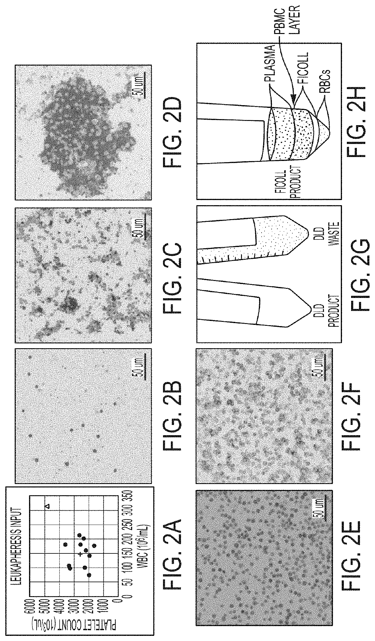
[0546]This study focuses on apheresis samples, which are integral to CAR-T-cell manufacture. The inherent variability associated with donor health, disease status and prior chemotherapy all impact the quality of the leukapheresis collection, and likely the efficacy of various steps in the manufacturing protocols (Levine, et al., Mol. Therapy: Meth. Clin. Dev. 4:92-101 (2017)). To stress test the automated DLD leukocyte enrichment, residual leukocytes (LRS chamber fractions) were collected from plateletpheresis donations which generally have near normal erythrocyte counts, 10-20-fold higher lymphocytes and monocytes and almost no granulocytes. They also have ˜10-fold higher platelet counts, as compared to normal peripheral blood.

[0547]12 donors were processed and yields were compared of major blood cell types and processivity by DLD versus Ficoll-Hypaque density gradient centrifugation, a “gold standard.” 4 of these donors were also assessed for “T-cell expansion capacity” over a 15-...

example 2

Add Back Experiment

[0582]Rationale

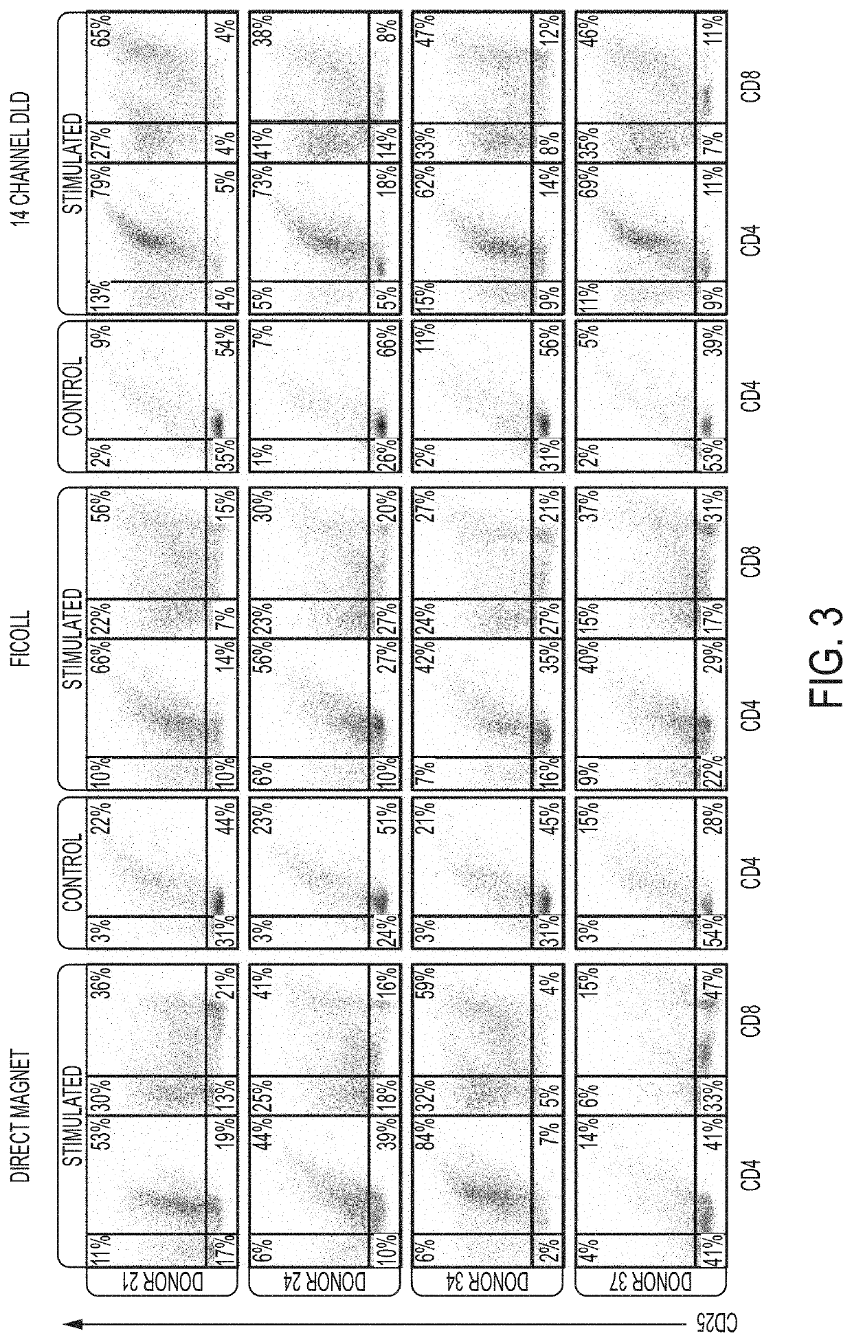
[0583]Previously, it has been found that WBC derived from the DLD isolation and purification are healthy and responsive to activation by CD3 / CD28 antibodies and differentiate towards their Tcm (T central memory) phenotype (Campos-Gonzalez, et al., SLAS, Jan. 23, 2018, published online doi.org / 10.1177 / 2472630317751214). Additionally, in the presence of IL-2 Tcm cells expand and proliferate accordingly and similarly to cells derived from other methods, like Ficoll.

[0584]A key feature of the DLD cell purification is the efficient removal of red blood cells and platelets to provide a highly purified white blood cells (WBC) product. In comparison, Ficoll-derived white blood cells (PBMC's) show more contaminating red blood cells and platelets depending on the sample quality. On average the platelet “contamination” in the Ficoll-derived cells is 44% has a range of about 22% of variability whereas the DLD cells exhibit only a 17% platelet contamination with...


PUM

PropertyMeasurementUnit
timeaaaaaaaaaa
sizeaaaaaaaaaa
diameteraaaaaaaaaa

Abstract

The present invention is directed to the use of microfluidics in the preparation of cells and compositions for therapeutic uses.

Description

CROSS REFERENCE TO RELATED APPLICATIONS[0001]This application is a continuation of U.S. Ser. No. 16 / 108,365, filed on Aug. 22, 2018, which claims the benefit of U.S. Provisional Patent Application No. 62 / 553,723, filed on Sep. 1, 2017; the benefit of U.S. Provisional Patent Application No. 62 / 567,553, filed on Oct. 3, 2017; the benefit of Provisional Patent Application No. 62 / 635,304, filed on Feb. 26, 2018; and the benefit of Provisional Patent Application No. 62 / 656,939, filed on Apr. 12, 2018; and which, in addition, is a continuation-in-part of PCT / US2017 / 057876, filed on Oct. 23, 2017. These prior applications are all incorporated by reference herein in their entireties.STATEMENT AS TO FEDERALLY SPONSORED RESEARCH[0002]This invention was made with government support under Grant No. CA174121 and No. HL110574 awarded by the National Institutes of Health. The government has certain rights in the invention.FIELD OF THE INVENTION[0003]The present invention is directed primarily to m...

Claims


Application Information

Patent Timeline
Patent Type & Authority Applications(United States)
IPC IPC(8): C12N5/0783C12N15/79B01L3/00A61K38/17A61K35/17A61P35/02
CPCC12N2533/54B01L3/502753C12N2531/00B01L2300/0864C12N2533/74C12N5/0636B01L2400/086B01L3/502761A61K35/17B01L2200/0652B01L2300/0816A61P35/02C12N5/0087
Inventor WARD, ANTHONYCAMPOS-GONZALEZ, ROBERTOSKELLEY, ALISONGANDHI, KHUSHROOCIVIN, CURTSTURM, JAMES C.GRISHAM, MICHAEL
Owner UNIV OF MARYLAND BALTIMORE
Why Eureka
Try Eureka