
Peptide nucleic acid conjugates
a nucleic acid and conjugate technology, applied in the field of conjugates, can solve the problems of limited tissue amount, limited clinical application of traditional gene expression analysis methods, and few efficient and reproducible methods that allow simultaneous and quantitative detection, and achieve the effect of minimizing interferen
- Summary
- Abstract
- Description
- Claims
- Application Information
AI Technical Summary
Benefits of technology
Problems solved by technology
Method used
Image

Examples
example 1
ody Conjugation
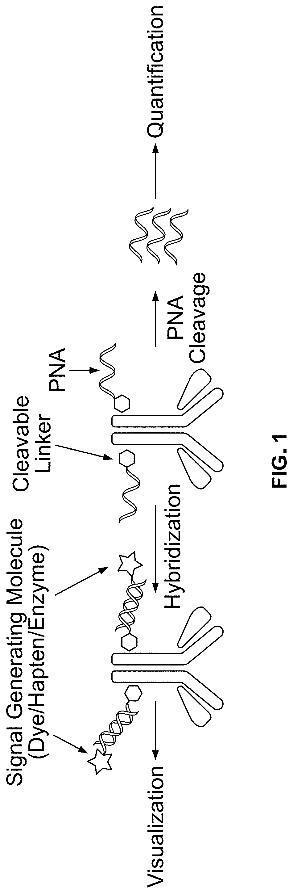
[0416]Exemplary conjugation of PNA to goat-anti-mouse (GAM), goat-anti-rabbit (GAR), and mouse-anti-DIG, is described below:
[0417](1) 100 micrograms of antibody (0.667 nmole) was diluted in PBS to 2 mg / mL and treated with 15 molar equivalent of SPDP-PEG-NHS (10 nmole from 10 mM DMSO stock solution) for 2 hours. SPDP-PEG-NHS was available from Quanta Biodesign, Ohio, USA (SPDP-dPEG8-NHS ester, product #10376).
[0418](2) The labeled antibody was purified with 7 KD MW cutoff Zeba spin desalting columns from ThermoFisher (product #89882).
[0419](3) 2 nmole PNA (8 uL from 251 μM PNA stock solution dissolved 1:1 (v / v) water DMF), was added to the antibody solution with the addition of 5 uL DMF, and 35 uL of PBS to yield approximately total of 100 uL of reaction solution. The mixture was incubated overnight at room temperature.
[0420](4) The conjugates were then purified using Zeba spin desalting columns were then used to purify PNA-conjugated antibodies from the reaction mixtu...
example 2
UV-Vis Measurement of Antibody-PNA Conjugates
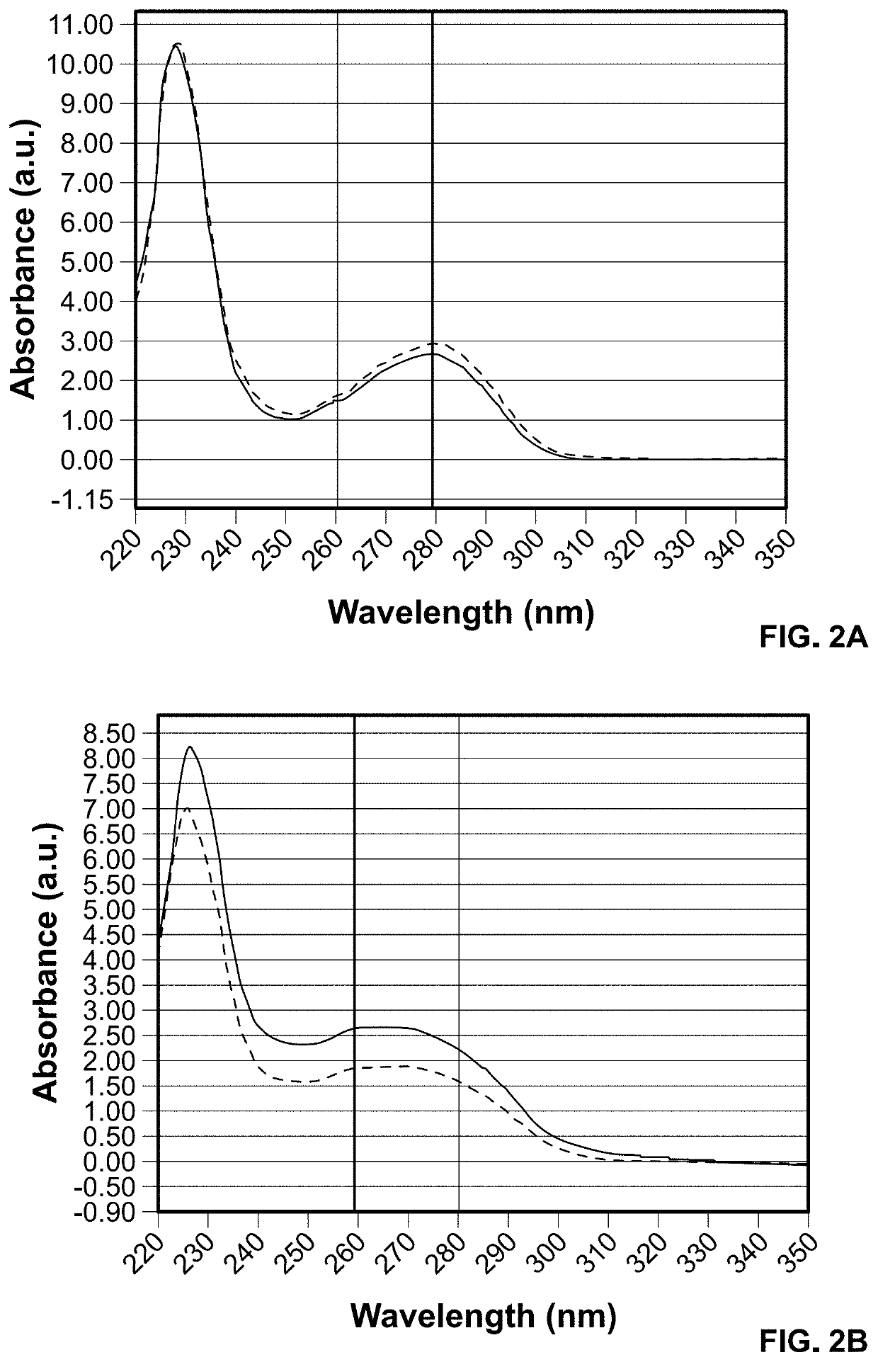
[0422]The PNA-conjugated antibodies were characterized with UV-Vis absorbance. The increase of absorbance at 260 nm in the antibody-PNA conjugates indicated successful incorporation of PNA (see FIG. 2). The ratio of A260 to A280 nm may be used to qualitatively assess the efficiency of conjugation and potentially estimate the number of PNA oligomers per antibody.
example 3
of PNA-Antibody Conjugate Using SA-HRP
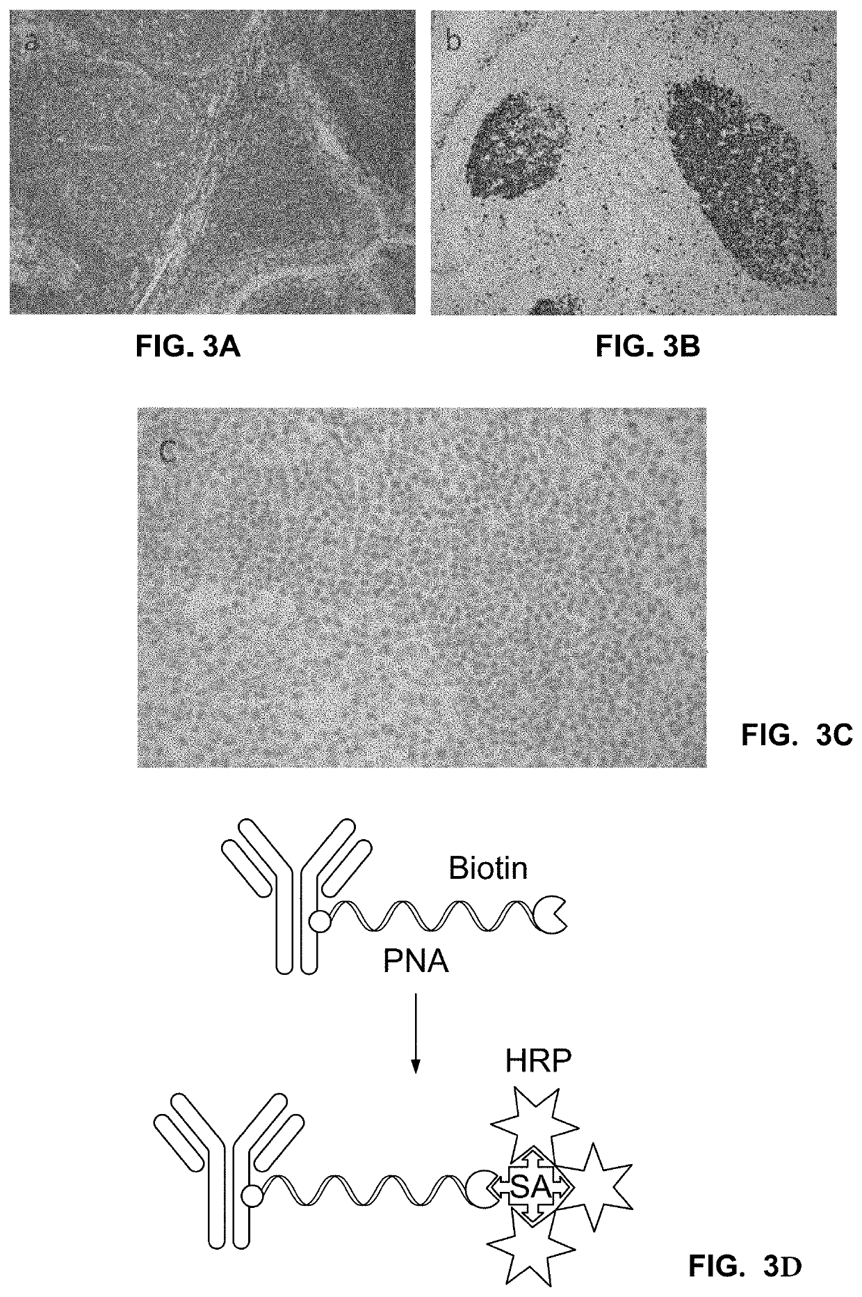
[0423]To prove that the conjugation of the short PNA (15 bases PNA, 10 bases PNA) oligo does not affect the binding affinity and specificity of the antibody, we performed IHC assays on tonsil tissues for different markers using PNA-conjugated secondary antibodies (primary and secondary). All IHC assays were run on Benchmark XT (Ventana) using protocols that were modified based on the specific assay. The PNA-conjugated antibodies were manually added as a titration at a volume of 100 uL.
[0424]The biotin label on the PNA tag was directly detected. Two tonsil slides were incubated with anti-CD45 and anti-Ki67 primary antibodies followed by incubation with PNA-conjugated GAR and GAM secondary antibodies, respectively. The slides were then incubated with Streptavidin-HRP (SA-HRP) followed by DAB deposition (chromogenic detection). The markers were observed at their expected locations (FIGS. 3A and 3B). No background signal was observed when primary an...
PUM
| Property | Measurement | Unit |
|---|---|---|
| Molar density | aaaaa | aaaaa |
| Molar density | aaaaa | aaaaa |
| Molar density | aaaaa | aaaaa |
Abstract
Description
Claims
Application Information