Unlock instant, AI-driven research and patent intelligence for your innovation.

X-ray imaging system and method for recognizing foreign object thereby

a technology of x-ray imaging and foreign objects, applied in the field of x-ray imaging systems and methods for recognizing foreign objects, can solve problems such as oversight, and achieve the effects of improving visual recognition level, higher screen resolution, and better visual recognition level

Pending Publication Date: 2022-04-14
SHIMADZU CORP
View PDF3 Cites 0 Cited by
  • Summary
  • Abstract
  • Description
  • Claims
  • Application Information

AI Technical Summary

Benefits of technology

The present invention aims to provide an X-ray imaging system and a method for recognizing foreign objects left inside a patient's body after surgery. The system and method improve visual recognition of foreign objects to prevent oversights and enable better identification of potential risks. The second display unit, which is separate from the X-ray imaging apparatus and connected via a network, provides a better visual recognition level for the foreign object image than the X-ray imaging apparatus' display. This makes it easier for users to visually recognize the foreign object and reduces the risk of complications.

Problems solved by technology

Nevertheless, an oversight may take place due to the reason of which a surgical instrument (foreign object) overlaps such as the used gauze sponge and a structure of the human body such as a bone even if it was confirmed that no foreign object of the surgical instrument such as the used gauze sponge is not left inside the patient (subject) body thereof by taking the X-ray image.

Method used


Image

  • X-ray imaging system and method for recognizing foreign object thereby
  • X-ray imaging system and method for recognizing foreign object thereby
  • X-ray imaging system and method for recognizing foreign object thereby

Examples


embodiment 1

Effect of Embodiment 1

[0077]The following effects can be obtained of Embodiment 1.

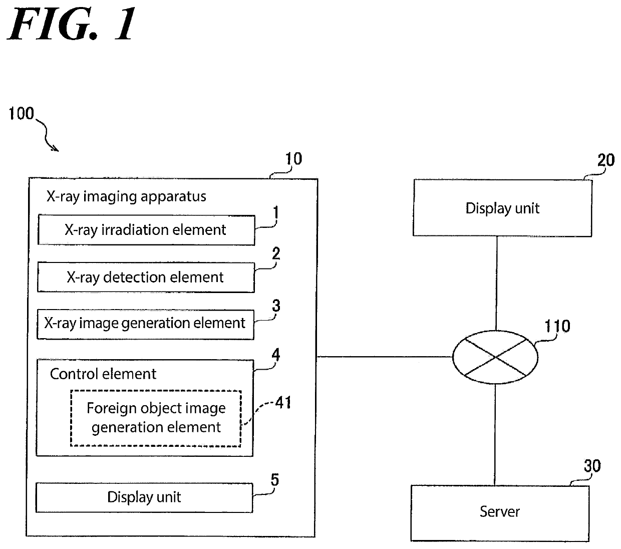
[0078]According to Embodiment 1, the X-ray imaging system 100 and the method for recognizing the foreign object generate the foreign object image 52 to recognize the foreign object 102 left inside the subject body 101 after the surgical operation. And the display unit 20 (second display unit) that display the foreign object image 52, which is installed separately from the X-ray imaging apparatus 10 and provides the better visual recognition level than the display unit 5 (first display unit) which the X-ray imaging apparatus 10 equips therewith. Therefore, the user can visually recognize the foreign object image 52 to recognize the foreign object 102 left inside the subject body 101 after the surgical operation on the display unit 20 that provides the better visual recognition level than the display unit 5 which the X-ray imaging apparatus 10 equips therewith. As results, the user easily recognizes the ...

embodiment 2

Effect

[0097]The following effects can be obtained according to Embodiment 2.

[0098]With regard to the X-ray imaging system 200 and the method for recognizing the foreign object according to Embodiment 2, the occurrence of the oversight of the foreign object 102, which is left inside the subject body 101 after the surgical operation, can be prevented.

[0099]Further, with regard to the X-ray imaging system 200 according to Embodiment 2, the effect below can be obtained due to the following structure.

[0100]With regard to the X-ray imaging system 200 according to Embodiment 2, the display unit 220 (second display unit) further comprises the first display 221 and the second display 222 positioned side by side on the wheeled platform monitor 40 which is parked in advance in the surgical operation room. And the foreign object image 52 is displayed on one display (e.g., the second display 222) of the first display 221 and the second display 222, and also the X-ray image 51 is displayed on the...

embodiment 3

[0103]Referring to FIG. 14 and FIG. 15, the inventors illustrate the structure of the X-ray imaging system 300 according to Embodiment 3 of the present invention. In addition, the same element as illustrated according to Embodiment 1 and Embodiment 2 has the same sign in FIG.

[0104]With regard to the X-ray imaging system 300 (referring to FIG. 14) according to t of Embodiment 3, the display unit 20 (referring to FIG. 14 and FIG. 15) installed and positioned in the surgical operation room, the display unit 220 installed on the wheeled platform monitor 40 parked therein in advance (referring to FIG. 14 and FIG. 15) can display the foreign object image 52. Specifically, with regard to the X-ray imaging system 300 according to Embodiment 3, any one of the display unit 20, the first display 221 and the second display 222 is capable of displaying the foreign object image 52 generated by the foreign object generation element 41.

[0105]In addition, the other structure and effect according to ...


PUM

PropertyMeasurementUnit
structureaaaaaaaaaa
Structureaaaaaaaaaa
voltageaaaaaaaaaa

Abstract

An X-ray imaging system has an X-ray imaging apparatus include a foreign object image generation element that generates a foreign object image to make sure there is no foreign object left inside a subject body after a surgical operation. A first display unit displays at least one image selected from a group consisting of an X-ray image and a foreign object image. The X-ray imaging system is connected to an X-ray imaging apparatus through a network and displays the foreign object image on the second display unit that provides a better visual recognition level than the first display unit.

Description

CROSS REFERENCE TO RELATED APPLICATIONS[0001]This application relates to, and claims priority from JP 2020-173585 filed Oct. 14, 2020, the entire contents of which are incorporated herein by reference.FIGURE SELECTED FOR PUBLICATION[0002]FIG. 1BACKGROUND OF THE INVENTIONField of the Invention[0003]The present invention relates to an X-ray imaging system and a method for recognizing a foreign object.Description of the Related Art[0004]Conventionally, a gauze counting device that counts the number of used gauze sponges that are used during a surgical operation is known to make sure whether the used gauze sponge is left inside a patient (subject) body or not. Such a gauze counting device is disclosed in JP 2018-48905 A.[0005]The gauze counting device disclosed in JP 2018-48905 A comprises an X-ray generation element that irradiates X-rays to a gauze sponge housing that houses a plurality of gauze sponges in which contrast strings are woven and an X-ray imaging element that images an X-...

Claims


Application Information

Patent Timeline
Patent Type & Authority Applications(United States)
IPC IPC(8): G01N23/04G06T7/00A61B6/00
CPCG01N23/04G06M11/00A61B6/465G06T7/0012G06T2207/10121G06T2207/30004A61B6/12A61B6/4405A61B6/464G01N2223/408G06T2207/10116
Inventor HOSOMI, NAOMASAHU, ERZHONGNAKAYA, TOMOHIRO
Owner SHIMADZU CORP