Imaging method using jointly a pet reconstruction and a compton reconstruction, preferably in 3D compton

a technology of compton and reconstruction, applied in the field of gamma ray source imaging, can solve the problems of large part of the photons not being detected correctly, short life of the radio-element used for the pet, and difficult to envisage the use of the device in the context of a surgical operation

Pending Publication Date: 2022-11-10
DAMAVAN IMAGING +1
View PDF1 Cites 0 Cited by
  • Summary
  • Abstract
  • Description
  • Claims
  • Application Information

AI Technical Summary

Benefits of technology

[0016]The present invention proposes a device, system and method for PET and COMPTON 3D imaging making it possible to overcome at least part of the drawbacks of the prior art.
[0019]Such an imaging method coupling the PET and at least one Compton camera allows the Compton image to be able to truncate the LORs (response lines) of the PET imaging with accuracy better than 2 cm.
[0045]According to another feature, said imager is coupled to a third imaging modality (CT-Scan or MRI, etc.) facilitating the fusion of images for a better diagnosis.

Problems solved by technology

However, the radio-elements used for the PET have a short life and are therefore often expensive.
However, in such configurations, a large part of the photons is not detected correctly because the probability of detection increases with the crossed scintillator thickness.
This configuration makes their use difficult to envisage in the context of a surgical operation.
Moreover, the PET scanners are very expensive (in the order of two million Euros) due to the volume of detectors required.
On the other hand, in the context of the preclinical imaging where the dimensions of the scanner are much smaller, the doses of radioactivity injected into the mice are very high, which can disrupt the physiological processes desired to be observed.
However, the applications of the Compton camera are still often limited today, in particular because of its cost, of the high level of noise on the images and of the difficulty in obtaining accurate reconstructions.
More generally in all of the technologies above, when scintillating crystals are used to produce an image of gamma radiation sources, the problem of the probabilistic nature of the gamma photon / matter interaction is encountered.
Furthermore, another general and constant problem in the field concerns the measurement of the time of arrival of the first photons on a given pixel, because it is important for the quality of the images obtained using the signals collected by the PET scanners and also for the temporal Compton cameras.
However, these crystals are very delicate to implement because they are extremely sensitive to atmospheric conditions (in particular hydrometry), which restricts their use in the form of monolithic crystals.
Nevertheless, the use of monolithic crystals is faced with a paradox with regard to the measurement of the time of arrival of the photons.
However, in a “pixelated” detector, the first photon detected by a given pixel is very likely to have undergone multiple reflections on the lateral faces of the crystal before being collected by the photodetector and the information on the real time of emission will therefore be inaccurate, if not totally lost.
Today, however, temporal resolutions measured with monolithic crystals are lower than those measured with pixel arrays.
Even more, the spatial resolution, signal / noise ratio of the PET imagers are limited by many factors: the thermalization of the positron, the non-collinearity of 511 keV photons, the structure of the detectors, the large proportion of the events recorded due to fortuitous coincidences, to the scattered pairs.
These unwanted events affect the measurement of the activity and increase the noise in the reconstructed images.
The time of flight information makes enhancements but is limited by the accuracy of the time measurement.
Moreover, the time of flight is today not very relevant on small scanners (for example brain scanner) or small animal imaging, the scanner being less than 30 centimeters in diameter.
However, due to the physical mechanisms of the emission of light in the scintillators, obtaining such low times of flight with crystals thick enough to be effective (approximately twenty millimeters) is no small challenge and progress is slow.

Method used


Image

  • Imaging method using jointly a pet reconstruction and a compton reconstruction, preferably in 3D compton
  • Imaging method using jointly a pet reconstruction and a compton reconstruction, preferably in 3D compton
  • Imaging method using jointly a pet reconstruction and a compton reconstruction, preferably in 3D compton

Examples


Embodiment Construction

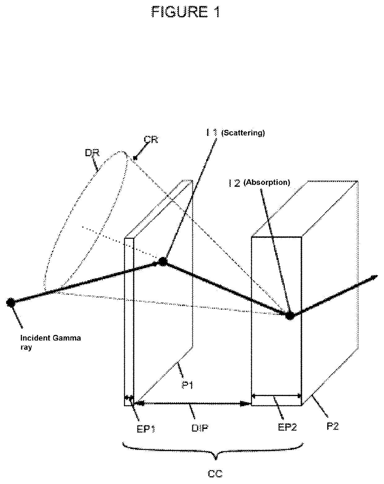
[0070]The present invention relates to an imaging system and method jointly using a PET-type coincidence reconstruction and a single-photon reconstruction at the same energy (511 keV) of the Compton 3D type. By the detection of gamma radiation implemented by a device including at least one PET type coincidence detection module. The two parts of the PET module are facing each other to make coincidence detections of pairs of photons. Said coincidence depends on a time window Δt (delta t) greater than the difference between the time of arrival t2 of the second photon detected by one of the PET cameras and that t1 of the first photon detected by the other PET camera (Δt=t2−t1). It's a system known as Time Of Flight (TOF).

[0071]In some embodiments, several pairs of PET cameras can be mounted facing each other forming a ring, arcs, etc. The purpose sought in these embodiments being that with these arrangements of PET cameras, it is possible to target large volumes and cover large surfaces...


PUM

PropertyMeasurementUnit
lengthsaaaaaaaaaa
thicknessaaaaaaaaaa
widthaaaaaaaaaa

Abstract

A PET and Compton imaging method implemented by a device including at least two facing PET modules. The device includes a Compton camera arranged outside a plane containing the PET modules for forming a trihedron with the PET modules and producing a Compton view. The acquisition fields of the PET and Compton views having an overlap area covering the object to be imaged. The device allowing the following steps to be carried out: acquisition of a Compton view; location of a dense area and its contour on the Compton view; Computation of the 2D map of the probability of detection of the presence of a source from the Compton view of the Compton camera; Coincidence detection by the PET cameras and association of a response line (LOR); and Segmentation of LORs crossing the dense area by using the detection probability determined by the Compton view.

Description

TECHNICAL FIELD[0001]The present application relates to the field of imaging and more particularly to the imaging of gamma ray sources. In particular, the invention relates to gamma ray detection imaging system and methods combining the Compton camera type detection and the Positron Emission Tomography (PET) type coincidence detection. The invention further relates to the use of the imaging and / or detection system in the fields in particular of astronomy, industry, in particular nuclear industry, and in the medical or veterinary fields.TECHNOLOGICAL BACKGROUND OF THE INVENTION[0002]Currently, the imaging of gamma ray sources (whose energy is generally greater than 30 KeV) is performed mainly for medical diagnostic purposes around three techniques: PET, SPECT and Compton Camera.[0003]The SPECT is based on scintigraphy and allows producing three-dimensional images and reconstructions of organs and their metabolism by means of a set of gamma cameras rotating around a patient. The SPECT...

Claims


Application Information

Patent Timeline
Patent Type & Authority Applications(United States)
IPC IPC(8): G01N23/20066
CPCG01N23/20066G01N2223/401G01T1/29G01T1/1603
Inventor ILTIS, ALAIN
Owner DAMAVAN IMAGING
Why Eureka
Try Eureka