Efficient single photon emission imaging

a single photon emission and computerized tomography technology, applied in tomography, instruments, applications, etc., can solve the problems of reducing the sensitivity of the collimator, requiring a long acquisition time, and a patient is subject to a relatively long period of discomfort, so as to enhance the image quality of the spect nuclear emission image, and shorten the data acquisition time

Inactive Publication Date: 2006-04-11
ULTRASPECT
View PDF11 Cites 69 Cited by
  • Summary
  • Abstract
  • Description
  • Claims
  • Application Information

AI Technical Summary

Benefits of technology

[0035]There is thus provided, in accordance with some embodiments of the present invention, a method of shortening the data acquisition time while at least maintaining and possibly enhancing image quality of a SPECT nuclear emission image acquired by a gamma camera.

Problems solved by technology

Sensitivity, however, is inversely related to geometric resolution, which means that improving collimator resolution (i.e., having smaller diameter holes) decreases collimator sensitivity, and vice versa.
SPECT is considered to be a very useful technique and a good tool for obtaining functional diagnostic information, however it requires the collection of large number of emitted photons (large statistics) and this means that in order to obtain the required number of photons, a long acquisition time is necessary.
Long acquisition time means that the patient is subjected to a relatively long period of discomfort, and, furthermore, the overall number of patients who can be imaged in a given time is relatively small—a feature that many medical institutes and hospitals regard as an extremely unfavourable and undesirable situation.
FBP algorithms suffer from image quality degradation when the number of angular projections is low or when the angular distribution is irregular.
Applying FBP algorithm on data set that includes high statistical noise causes degradation to the quality of the reconstructed image.
The current use of collimators results in a rather low detection efficiency of conventional SPECT, which leads to a prolonged data acquisition time and the need to administer high dosage of the radiopharmaceutical substance.
The collimators used in standard size gamma camera are heavy and costly.
Image quality is strongly adversely affected by that noise.
However, the resolution of the image is limited by the size of the pixels, so that larger matrices are desirable in situations where small features in the image have to be resolved.
This attenuation causes degradation of image quality due to loss of photons and more importantly, image distortion due to varying amount of attenuation between different organs of the body and the detector as it views the body from various direction.
Since attenuation through the dense parts of the body is considerably high the number of transmitted photons is low and thus image quality of the reconstructed attenuation map is limited by the time spent on acquiring the transmission data.

Method used


Image

  • Efficient single photon emission imaging
  • Efficient single photon emission imaging
  • Efficient single photon emission imaging

Examples


Embodiment Construction

[0057]The general objective of the present invention, which will be described subsequently in greater detail, is to provide a novel technique for acquisition and reconstruction of SPECT single photon emission images in which the duration of the time consuming data acquisition step is substantially reduced (with respect to known techniques), thus decreasing patient discomfort, potentially improving image quality and increasing the Gamma camera's throughput.

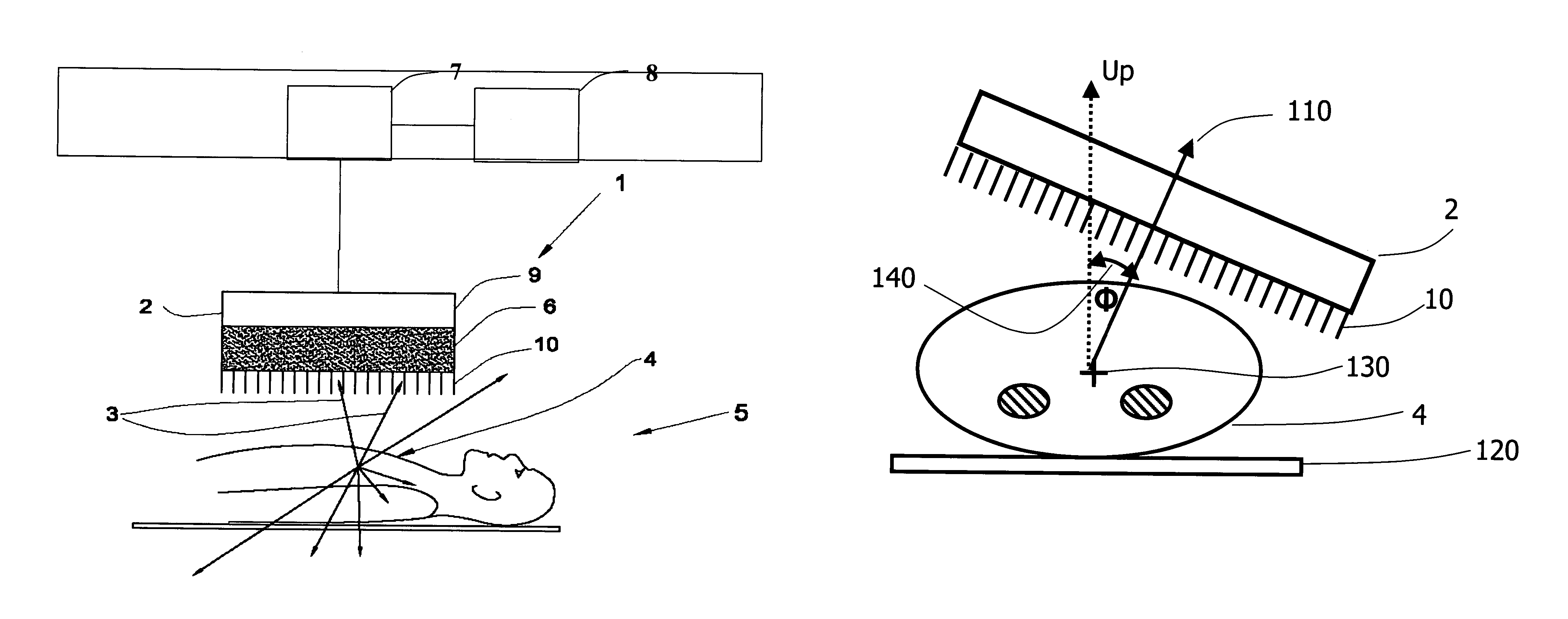
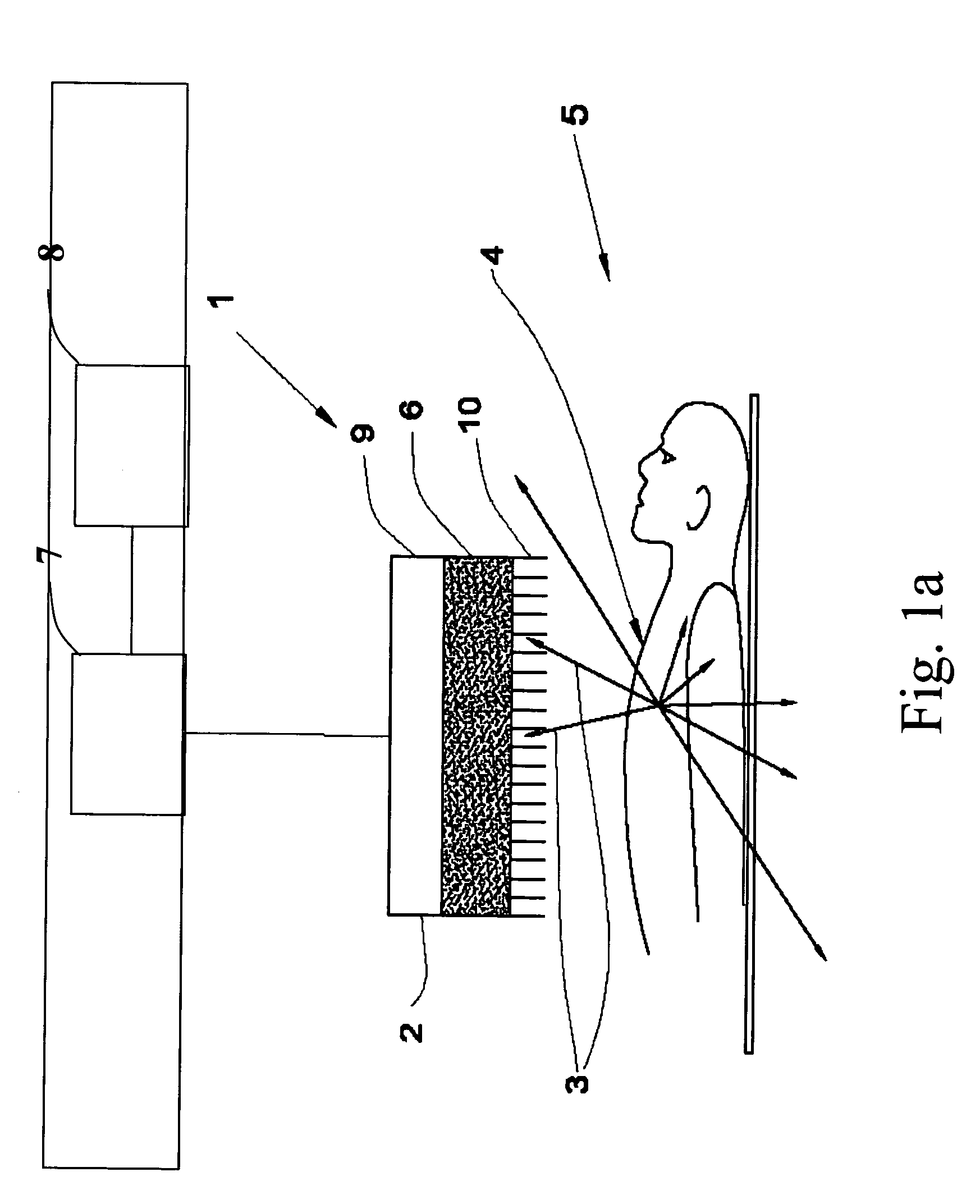
[0058]Reference is first made to FIG. 1a depicting a side view of a simplified schematic diagram of gamma camera in accordance with the present invention, for obtaining a SPECT image of a portion of a body that has been administered by a radiopharmaceutical substance radiating gamma rays.

[0059]The gamma camera 1 comprises a detector 2 mounted above an inspected portion 4 of a body 5, position logic circuitry 7 and a data analysis computer 8, all connected appropriately.

[0060]Detector 2 includes at least one photon detector crystal ...


PUM


Abstract

A method of diagnostic imaging in a shortened acquisition time for obtaining a reconstructed diagnostic image of a portion of a body of a human patient who was administered with dosage of radiopharmaceutical substance radiating gamma rays, using SPECT. The method comprises acquiring photons emitted from said portion of the body, by means of a detector capable of converting the photons into electric signals, wherein the total time of photon acquiring is substantially shorter than the clinically acceptable acquisition time; processing said electric signals by a position logic circuitry and thereby deriving data indicative of positions on said photon detector crystal, where the photons have impinged the detector; andreconstructing an image of a spatial distribution of the pharmaceutical substance within the portion of the body by iteratively processing said data.

Description

FIELD OF THE INVENTION[0001]The present invention relates to nuclear medicine imaging More particularly it relates to an efficient method for single photon emission computerized tomography imaging.BACKGROUND OF THE INVENTION[0002]Nuclear Medicine imaging techniques enable to acquire functional information on a patient's specific organ or body system. This functional information is attained from analysis of internal radiation from pharmaceutical substance administered to the patient, which is labelled with a radioactive isotope. The radioactive isotope decays, resulting in the emission of gamma rays, thus providing information on the concentration of the radiopharmaceutical substance in regions of the patient's body. An instrument for the detection of gamma ray emissions of the radiopharmaceutical substance administered in the body is known as gamma camera. The Gamma camera collects gamma ray photons that are emitted from the patient's body, and the collected data is used to reconstr...

Claims


Application Information

Patent Timeline
Patent Type & Authority Patents(United States)
IPC IPC(8): G01T1/166A61B6/00G01T1/164G06T11/00
CPCA61B6/4057G06T11/006A61B6/037G01T1/1644A61B6/503A61B6/541A61B6/583G06T2211/424
Inventor OAKNIN, JACOBSHWARTZ, SHOULAMIT C.OHANA, ISRAEL
Owner ULTRASPECT
Why Eureka
Try Eureka