Composition and method for the repair and regeneration of cartilage and other tissues

a technology applied in the field of cartilage and other tissues, to achieve the effect of improving coagulation/solidification

Inactive Publication Date: 2006-12-12
SMITH & NEPHEW ORTHOPAEDICS
View PDF36 Cites 214 Cited by
  • Summary
  • Abstract
  • Description
  • Claims
  • Application Information

AI Technical Summary

Benefits of technology

[0024]Also in accordance with the present invention, there is provided a method for repairing a tissue of a patient, said method comprising the step of introducing into said tissue a temperature-dependent polymer gel composition such that said composition adhere to the tissue and promote support for cell proliferation for repairing the tissue.
[0034]The blood component can also comprise an anticoagulant such as citrate, heparin or EDTA. To the opposite the blood component can comprise a pro-coagulant such as thrombin, calcium, collagen, ellagic acid, epinephrine, adenosine diphosphate, tissue factor, a phospholipid, and a coagulation factor like factor VII to improve coagulation / solidification at the site of introduction.

Problems solved by technology

Some of these techniques have achieved a certain level of acceptance in clinical practice but this has mainly been so due to the absence of any practical and clearly effective method of improving the repair response compared to that found when the family of bone marrow stimulation techniques is applied.
One main obstacle towards the development of an effective cartilage repair procedure is the absence of a composition and method to provide an appropriate macromolecular environment within the space requiring cartilage growth (cartilage defect or other site requiring tissue bulking or reconstruction) This macromolecular environment or matrix should 1) be amenable to loading with active biological elements (cells, proteins, genes, blood, blood components) in a liquid state 2) then be injectable into the defect site to fill the entire defect or region requiring cartilage growth 3) present a primarily nonproteinaceous environment to limit cell adhesion and cell-mediated contraction of the matrix, both of which induce a fibrocytic cellular phenotype (fibrous tissue producing) rather than chondrocytic cellular phenotype (cartilaginous tissue producing) and which can also disengage the matrix from the walls of the defect 4) be cytocompatible, possessing physiological levels of pH and osmotic pressure and an absence of any cytotoxic elements 5) be degradable but present for a sufficiently long time to allow included biologically active elements to fully reconstitute a cartilaginous tissue capable of supporting mechanical load without degradation.

Method used


Image

  • Composition and method for the repair and regeneration of cartilage and other tissues
  • Composition and method for the repair and regeneration of cartilage and other tissues
  • Composition and method for the repair and regeneration of cartilage and other tissues

Examples


example 1

Mixing of Thermogelling Chitosan Solution with Primary Chondrocytes for In Vitro Growth of Cartilage

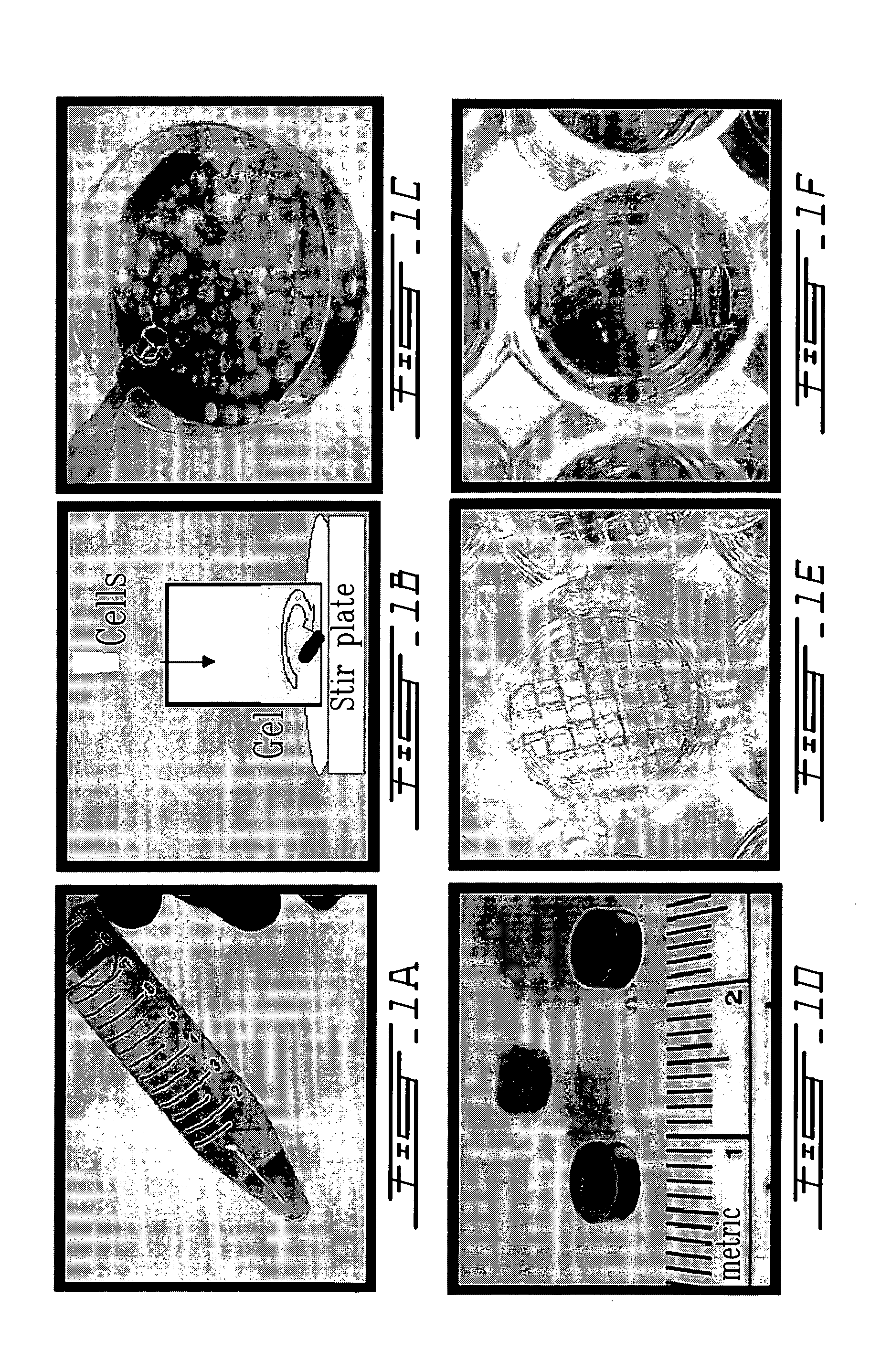
[0079]Chitosan (0.22 g, 85% deacetylated) as an HCl salt powder was sterilized by exposure to ultraviolet radiation in a biological laminar flow hood and then dissolved in 7.5 ml H2O resulting in a pH near 5.0. D(+)-glucosamine (0.215 g, MW 215.6) was dissolved in 10 ml of 0.1M NaOH and filter sterilized using a 22 μm pore size disk filter. Glycerol phosphate (0.8 g, MW 297 including 4.5 mole water per mole glycerol phosphate) was dissolved in 2.0 ml of H2O and filter sterilized using a 22 μm pore size disk filter. 2.25 ml of the glucosamine solution was added drop-by-drop under sterile conditions to the chitosan solution with agitation at a temperature of 4° C. Then 1 ml of the glycerol phosphate solution was added under the same conditions. This final solution is still a liquid and remains so for an extended period (i.e. days) if the temperature is kept low, i.e. near 4° C. The pH o...

example 2

Mixing of Thermogelling Chitosan Solution with Primary Chondrocytes and Subcutaneous Injection for In Vivo Growth of Cartilage

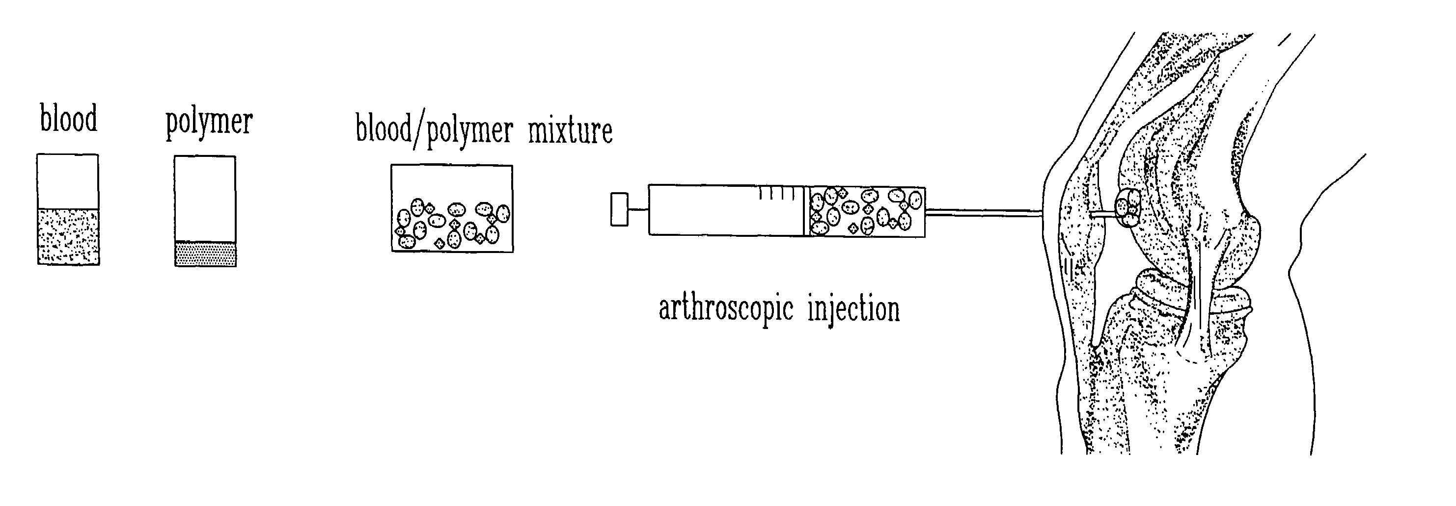
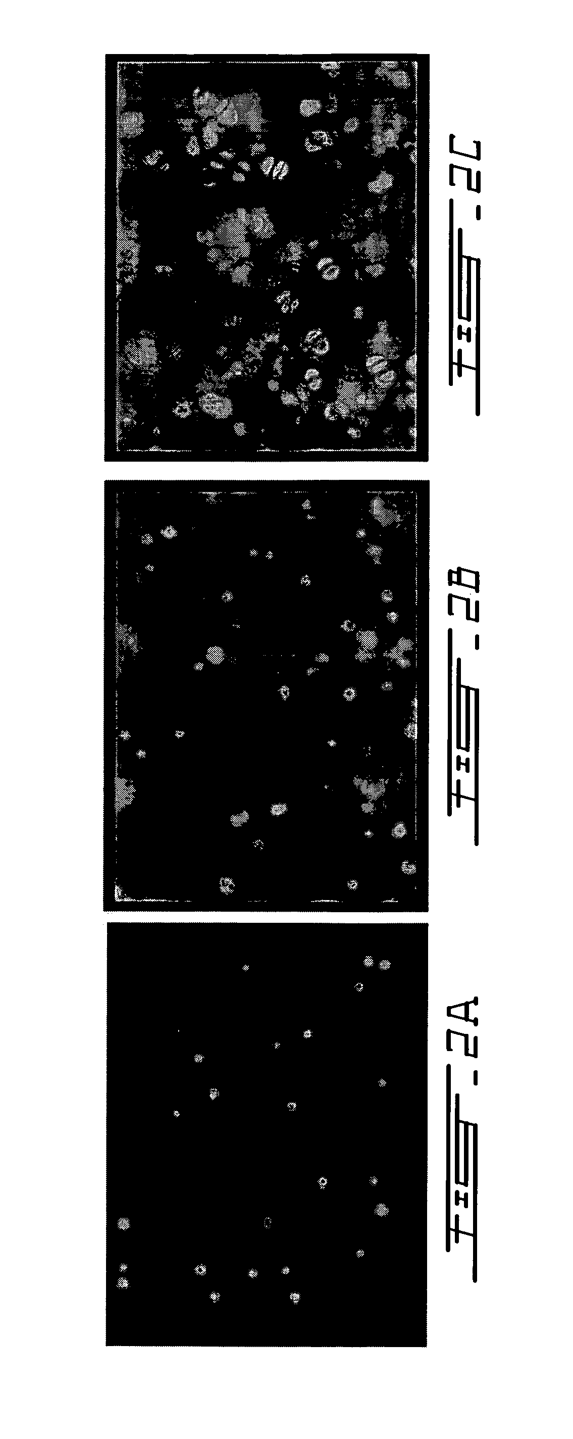
[0087]To demonstrate that this in situ gelling system can be employed in animals, athymic mice (CD1 nu / nu) were subjected to dorsal, subcutaneous injections of 100 to 300 μl of chitosan gel described in Example 1, containing 10 million calf articular chondrocytes per ml (FIG. 7). A cell pellet of primary calf chondrocytes was admixed with liquid chitosan gel at 4° C. to achieve a concentration of 1 to 2×10⁷ cells / ml, and injected in liquid form as 100 μl subcutaneous dorsal implants in anesthetized nude mice. In situ gelling was apparent by palpation 5 to 10 minutes post-injection.

[0088]Control mice were similarly injected with chitosan gel alone. A palpable gel was formed within 10 minutes of injection. Implants were recovered at 21, 48, and 63 days post-injection. Toluidine blue staining revealed the gross production of GAG-rich extracellular matrix by the ...

example 3

Adhesion of Thermogelling Chitosan Solution to Cartilage and Bone Surfaces

[0093]One of the most significant advantages of this chitosan thermogelling formulation for cartilage repair is its ability to conform and adhere to irregular cartilage defects and other irregularly shaped cavities in the body that require tissue repair, regeneration, reconstruction or bulking. Many current tissue repair procedures suffer drastically in this respect. Chitosan-glycerol phosphate liquid gel without cells was delivered ex vivo to porcine femoral condylar intra-chondral (not involving bone) defects. Disc-shaped defects in the articular cartilage were created using a biopsy punch (FIG. 12A) and the chitosan solution described in Example 1 was injected into these defects and allowed to solidify in an incubator at 37° C. The articulating cartilage surface was opposed and simulated joint motions were performed after which the gel was observed to remain in the cartilage defect (FIG. 12B). The gel not o...


PUM

PropertyMeasurementUnit
molecular weightaaaaaaaaaa
pore sizeaaaaaaaaaa
temperatureaaaaaaaaaa

Abstract

The present invention relates to a new method for repairing human or animal tissues such as cartilage, meniscus, ligament, tendon, bone, skin, cornea, periodontal tissues, abscesses, resected tumors, and ulcers. The method comprises the step of introducing into the tissue a temperature-dependent polymer gel composition such that the composition adhere to the tissue and promote support for cell proliferation for repairing the tissue. Other than a polymer, the composition preferably comprises a blood component such as whole blood, processed blood, venous blood, arterial blood, blood from bone, blood from bone-marrow, bone marrow, umbilical cord blood, placenta blood, erythrocytes, leukocytes, monocytes, platelets, fibrinogen, thrombin and platelet rich plasma. The present invention also relates to a new composition to be used with the method of the present invention.

Description

CROSS-REFERENCE TO RELATED APPLICATIONS[0001]This application is a continuation of U.S. Ser. No. 09 / 896,912 filed Jun. 29, 2001 now abandoned, which claims the benefit under 35 U.S.C. § 119(e) of United States Provisional Application Nos. 60 / 214,717 filed Jun. 29, 2000.BACKGROUND OF THE INVENTION[0002](a) Field of the Invention[0003]The invention relates to a composition and method of application to improve the repair and to regenerate cartilaginous tissues and other tissues including without limitation meniscus, ligament, tendon, bone, skin, cornea, periodontal tissues, abscesses, resected tumors, and ulcers.[0004](b) Description of Prior Art1) The Cartilage Repair Problem:Cartilage: Structure, Function, Development, Pathology[0005]Articular cartilage covers the ends of bones in diarthroidial joints in order to distribute the forces of locomotion to underlying bone structures while simultaneously providing nearly frictionless articulating interfaces. These properties are furnished ...

Claims


Application Information

Patent Timeline
Patent Type & Authority Patents(United States)
IPC IPC(8): A01N43/04A01N47/08A61D99/00A61F2/00A61K38/02A61L27/00A61F2/10A61F2/14A61F2/28A61L27/18A61L27/20A61L27/22A61L27/38A61L27/50
CPCA61K31/727A61K31/728A61K31/737A61K35/15A61K35/16A61K35/18A61K35/19A61K38/363A61K38/4833A61K38/4846A61L27/18A61L27/20A61L27/22A61L27/3616A61L27/3641A61L27/3687A61L27/3691A61L27/3804A61L27/3839A61L27/50A61K9/0019A61K9/0024A61K47/10A61K9/06A61K47/36A61K2300/00A61P19/04A61L27/38
Inventor HOEMANN, CAROLINE D.BUSCHMANN, MICHAEL D.MCKEE, MARC D.
Owner SMITH & NEPHEW ORTHOPAEDICS
Why Eureka
Try Eureka