X-ray diagnostic device for mammography examinations

a diagnostic device and mammography technology, applied in the field of x-ray diagnostic devices for mammography examinations, can solve the problems of unnecessarily high x-ray dose, and achieve the effect of reducing the risk of exposing the patient and good x-ray image quality

Active Publication Date: 2007-04-03
SIEMENS HEALTHCARE GMBH
View PDF3 Cites 7 Cited by
  • Summary
  • Abstract
  • Description
  • Claims
  • Application Information

AI Technical Summary

Benefits of technology

[0006]An object of an embodiment of the invention is to create an X-ray diagnostic unit having an arrangement that reduces the risk of exposing the patient to be examined to an unnecessarily high dose of X-ray radiation.
[0007]An object may be achieved according to an embodiment of the invention by providing the object table with a sensing arrangement that is connected to a transport device for the X-ray grid. When applying the attachment unit to the object table, the attachment unit preferably actuates the sensing arrangement in such a way that the transport device transports the X-ray grid out of the radiation field. The automatic transportation of the X-ray grid out of the X-ray field when the magnification table or the biopsy unit is applied to the object table ensures that the patient receives only the minimum X-ray dose that is necessary to obtain a good X-ray image quality.

Problems solved by technology

If an operator forgets to move the X-ray grid out of the X-ray field when using magnification or when performing a biopsy examination, an unnecessarily high X-ray dose can accidentally be given to the breast to be examined in order to obtain a good image quality, because the X-ray grid absorbs a large proportion of the X-ray dose.

Method used


Image

  • X-ray diagnostic device for mammography examinations
  • X-ray diagnostic device for mammography examinations
  • X-ray diagnostic device for mammography examinations

Examples


Embodiment Construction

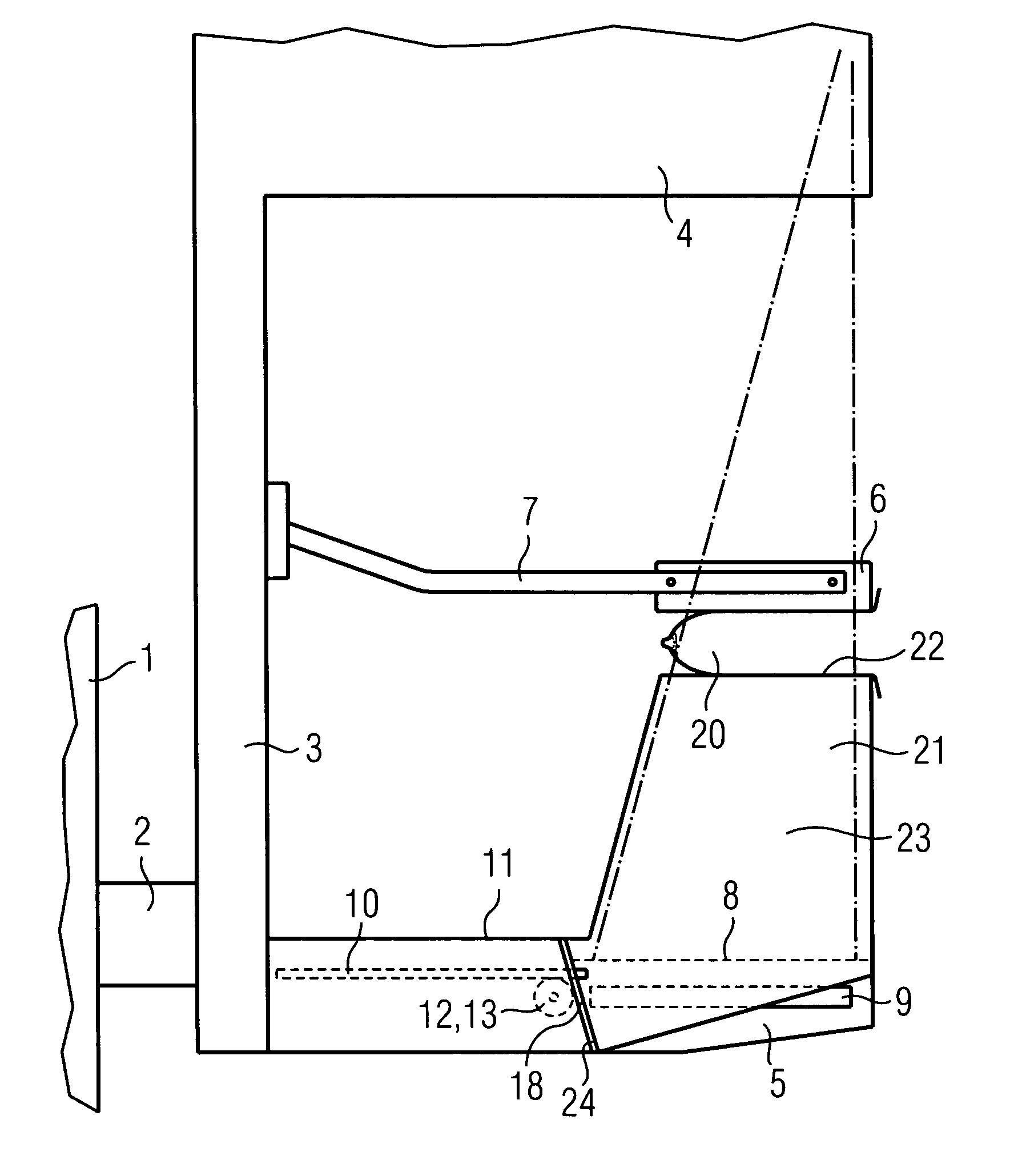
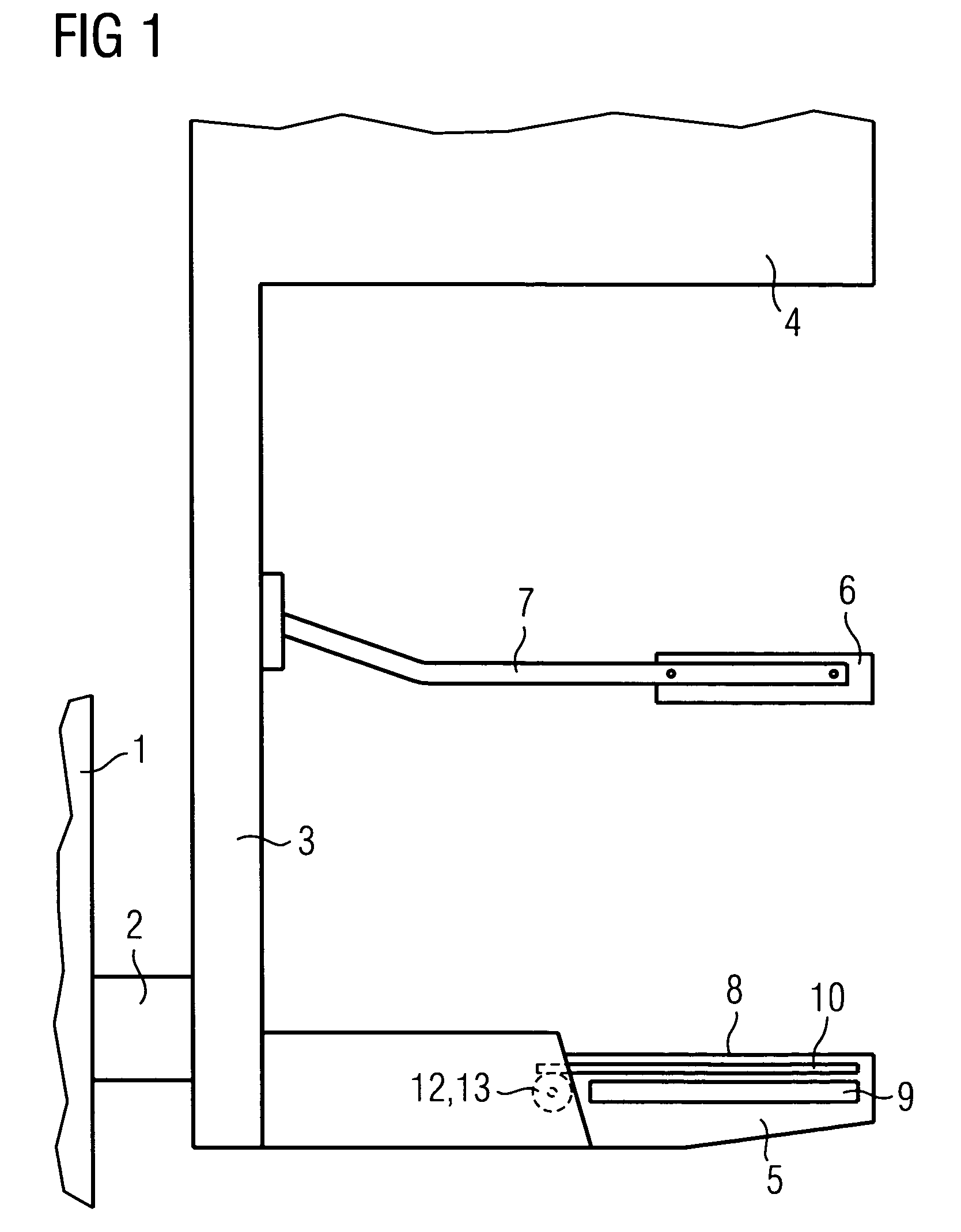
[0012]FIG. 1 shows a side view of a section of an X-ray diagnostic unit for mammography examinations. A device stand 1 is shown in FIG. 1 that is connected via a shaft 2 to an arm 3 for an X-ray tube and an object table 5. A compression plate 6 is arranged between the X-ray tube 4 and the object table 5 and is connected via a bracket 7 to the arm 3 along which it can move.

[0013]The object table 5 includes a table surface 8, an image receptor 9 and an X-ray grid 10 arranged between the table surface 8 and the image receptor 9. A transport device for the X-ray grid 10 is also located in the object table 5 and is shown in FIG. 2 and described in more detail with reference to this figure.

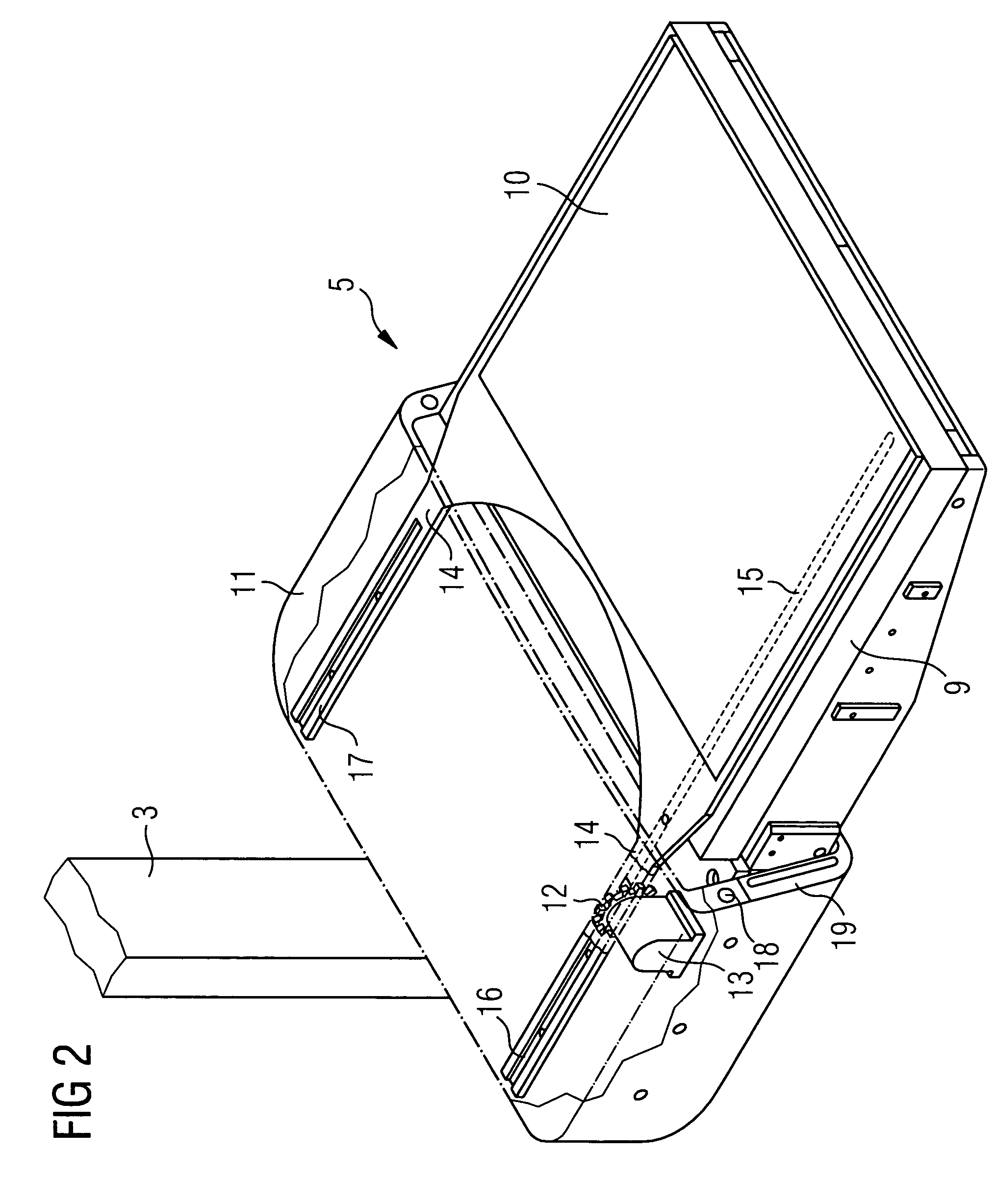
[0014]FIG. 2 shows a perspective view of the object table 5. The table surface 8 (FIG. 1) has been removed in the figure to provide a clear view of the X-ray grid 10. A housing 11 for the object table 5 has been indicated simply by dashed lines to provide a clearer view of the transport device mentioned...


PUM


Abstract

An X-ray diagnostic device is for mammography examinations and includes an arm for an X-ray tube and an object table having an image receptor. The X-ray tube is able to emit a radiation field towards the image receptor. Further, an X-ray grid is included, that is introducable into the radiation field, as well as an attachment unit that can be attached to the object table. In order to obtain an X-ray diagnostic unit having an arrangement that eliminates the risk of exposing the patient to be examined to an unnecessarily high dose of X-ray radiation, the object table is provided with a sensing arrangement that is connected to a transport device for the X-ray grid. Upon applying the attachment unit to the object table, the attachment unit actuates the sensing arrangement in such a way that the transport device transports the X-ray grid out of the radiation field.

Description

[0001]The present application hereby claims priority under 35 U.S.C. §119 on German patent application number DE 103 37 936.3 filed Aug. 18, 2003, the entire contents of which are hereby incorporated herein by reference.FIELD OF THE INVENTION[0002]The invention generally relates to an X-ray diagnostic device for mammography examinations. Preferably, it relates to a device having an arm for an X-ray tube and having an object table having an image receptor. The X-ray tube is preferably able to emit a radiation field towards the image receptor. The device further preferably includes an X-ray grid, an attachment unit that can be attached to the object table, and a compression plate, arranged between the X-ray tube and the object table, which is connected to the arm and can be moved along the arm.BACKGROUND OF THE INVENTION[0003]A brochure from the Siemens company entitled “MAMMOMAT 3000 Nova” shows an X-ray diagnostic unit for mammography examinations. The attachment unit mentioned in t...

Claims


Application Information

Patent Timeline
Patent Type & Authority Patents(United States)
IPC IPC(8): A61B6/04A61B6/00
CPCA61B6/0414A61B6/4283A61B6/502A61B6/4291
Inventor ANDREASSON, JESPERKARLSSON, STEFAN
Owner SIEMENS HEALTHCARE GMBH
Why Eureka
Try Eureka