Methods and devices for improving percutaneous access in minimally invasive surgeries

a technology for minimally invasive surgery and percutaneous access, applied in the direction of prosthesis, catheter, osteosynthesis device, etc., can solve the problems of prolonged recovery time, increased risk of infections, and increased technical difficulty of these operations, so as to reduce the risk of iatrogenic injuries to vital structures, improve visualization, and reduce the effect of technical difficulty

Active Publication Date: 2005-03-24
STRYKER EURO OPERATIONS HLDG LLC
View PDF99 Cites 561 Cited by
  • Summary
  • Abstract
  • Description
  • Claims
  • Application Information

AI Technical Summary

Benefits of technology

[0015] Among the advantages of this invention may be one or more of the following. The invention provides novel devices and methods for improving percutaneous surgeries for all applications and approaches in the body that previously required open surgery. These improvements will be beneficial to both patients and surgeons in that this invention will reduce the technical difficulty of these operations, improve visualization, decrease risks of iatrogenic injuries to vital structures, decrease length of hospitalization and associated costs, decrease operative time, decrease recovery time, and decrease postoperative pain. This invention provides the ability to adjust the length of the minimal access portals either inside or outside the patient to account for the varying depth of the pathology within the body. The graduated markers in millimeter increments etched on the outside of the cannula allow a determination of the depth of the pathology relative to the skin thus allowing the user to make adjustments to the fixation points such that they are aligned to the same depth. The invention further allows fixing two points percutaneously along the lateral aspect of the spine by directly placing the connecting device between the fixation points without visualizing the entire connecting device or by coming in along an arc or using fluoroscopic imaging. The invention also provides the ability to sequentially connect a fixation device percutaneously between more than two points simultaneously and only directly visualizing the fixation points and not the entire connecting device. This scaleable feature is currently a major limitation of other minimal access devices. Another advantage of this invention is the ability to perform a direct approach to the fixation of two or more points with the option to place the connecting device beneath the lumbodorsal fascia or through an incision, created by instruments in the lumbodorsal fascia between each fixation points rather than being confined to go beneath the fascia between the fixation points or through a larger opening in the fascia which requires greater tissue expansion and results in greater postoperative pain. The invention also provides a device that easily connects the portals at the sequential fixation points and simultaneously delivers objects such as connecting devices, or tools between the fixation points even if the fixation points are not in a perfectly straight line. The invention also provides the ability to lock the working cannula to the pedicle screw and is the first pedicle screw to feature an appendage for connecting a percutaneous device. It is the first device to allow easy retrieval of a medical device in contrast to the other systems that are designed for insertion of medical devices without a method or features that are designed for retrieval of the device. The cannulae can be easily removed from the fixation points and have the ability to reconnect to the fixation points and remove the connecting device even after having completed the surgery or after connecting the device to the fixation points and removing the cannulae. The invention also provides a side-working channel in addition to the central working channel to allow easier placement of connecting devices between fixation points without obstructing the visual portal as in other devices with only a single working channel that also doubles as a visualization channel. The invention also provides a fixed or rotating apparatus at various positions on any of the working channels or portals that can be used for optics, illumination, irrigation, or aspiration or combination thereof. The invention also provides a carrier device for carrying devices to be connected at the fixation points of the cannulae. This carrier device may be made of a material that is stiff, malleable, flexible or expandable at body temperature. By being malleable this carrier device may be used as a template on the skin surface between the cannulae prior to insertion and will be particularly beneficial when the cannulae do not line up in a straight line. The present invention has applications in a wide range of surgical procedures, and in particular in spinal procedures such as laminotomy, laminectomy, foramenotomy, facetectomy and discectomy, fusions or disc replacements using an anterior, posterior, postero-lateral, or a lateral approach to the disc space, facet, laminas, pedicles, or transverse processes. The devices and instruments of the present invention have application to surgical techniques that permit each of these several types of surgical procedures to be performed via a single or multiple sequential working channels. The present invention also has application to surgical techniques for preparing a disc space for insertion of an implant into the disc space.

Problems solved by technology

These procedures result in extensive tissue traumatization and consequently in prolonged recovery time, risk of infections, high hospitalization costs, pain that can be more severe than the pain due to the initial ailment, and in some cases permanent scarring.
The avoidance of general anesthesia reduces post-operative recovery time and the risk of complications.
Minimally invasive surgical procedures are especially desirable for spine surgeries because spine pathologies are located deep within the body without clear muscle planes and there is danger of damaging the adjacent neural and vascular tissues.
The destruction to the spinal structures is even more extensive during fusion procedures, which require more lateral tissue dissection and exposure to access the transverse processes and pedicles for placement of pedicle screws, rod constructs for stability, and bone graft under direct vision.
Unfortunately, the effectiveness and safety of the enzyme, chymopapain used for chemonucleolysis, have been complicated by severe spasms, post-operative pain, and sensitivity reactions including anaphylactic shock.
Loupe magnification and microscopes are helpful for improving visualization but are not effective without retractor systems and specialized instruments and devices to make minimal access surgery effective.
A problem that occurs frequently in minimally invasive surgical procedures is related to the fact that it is not always known how deep the pathology is located.
Placement of these connecting elements requires open surgery, which is currently one of the major limitations of other percutaneous cannula access methodologies.

Method used


Image

  • Methods and devices for improving percutaneous access in minimally invasive surgeries
  • Methods and devices for improving percutaneous access in minimally invasive surgeries
  • Methods and devices for improving percutaneous access in minimally invasive surgeries

Examples


Embodiment Construction

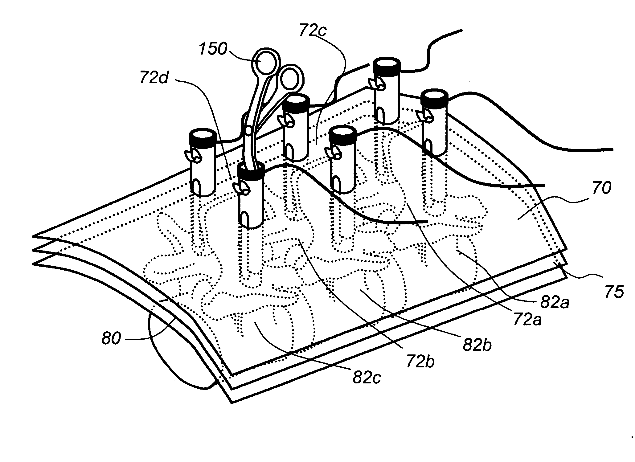
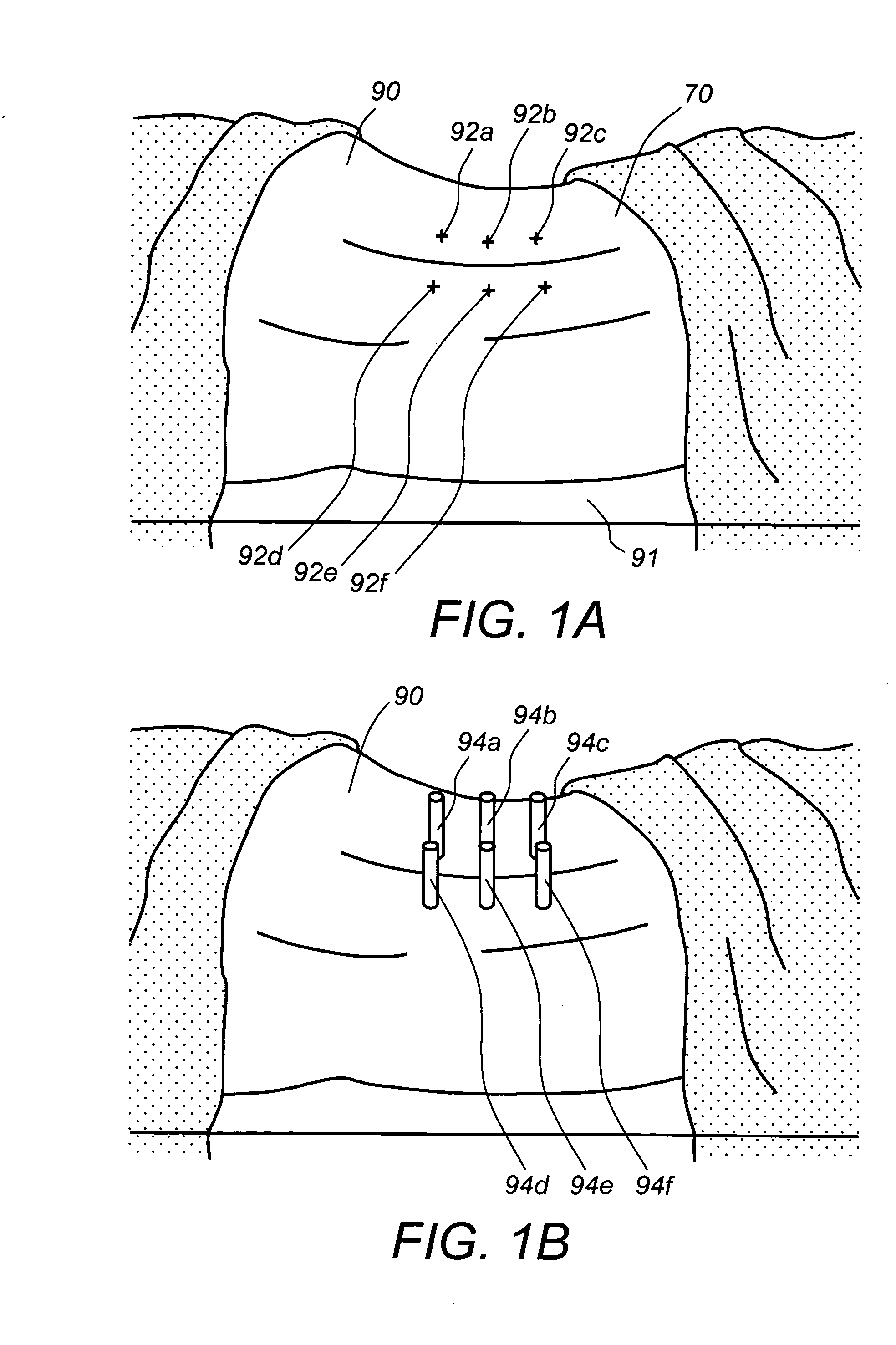
[0049] Referring to FIG. 1A, a patient 90 is positioned prone, lying flat on an operating table 91 in preparation for a minimally invasive surgery (MIS). Locations 92a-92f are marked on the patient's lower back corresponding to pedicle locations of adjacent vertebrae. For MIS procedures portals 94a-94f are inserted through skin incisions performed in the marked locations 92a-92f, respectively, shown in FIG. 1B.

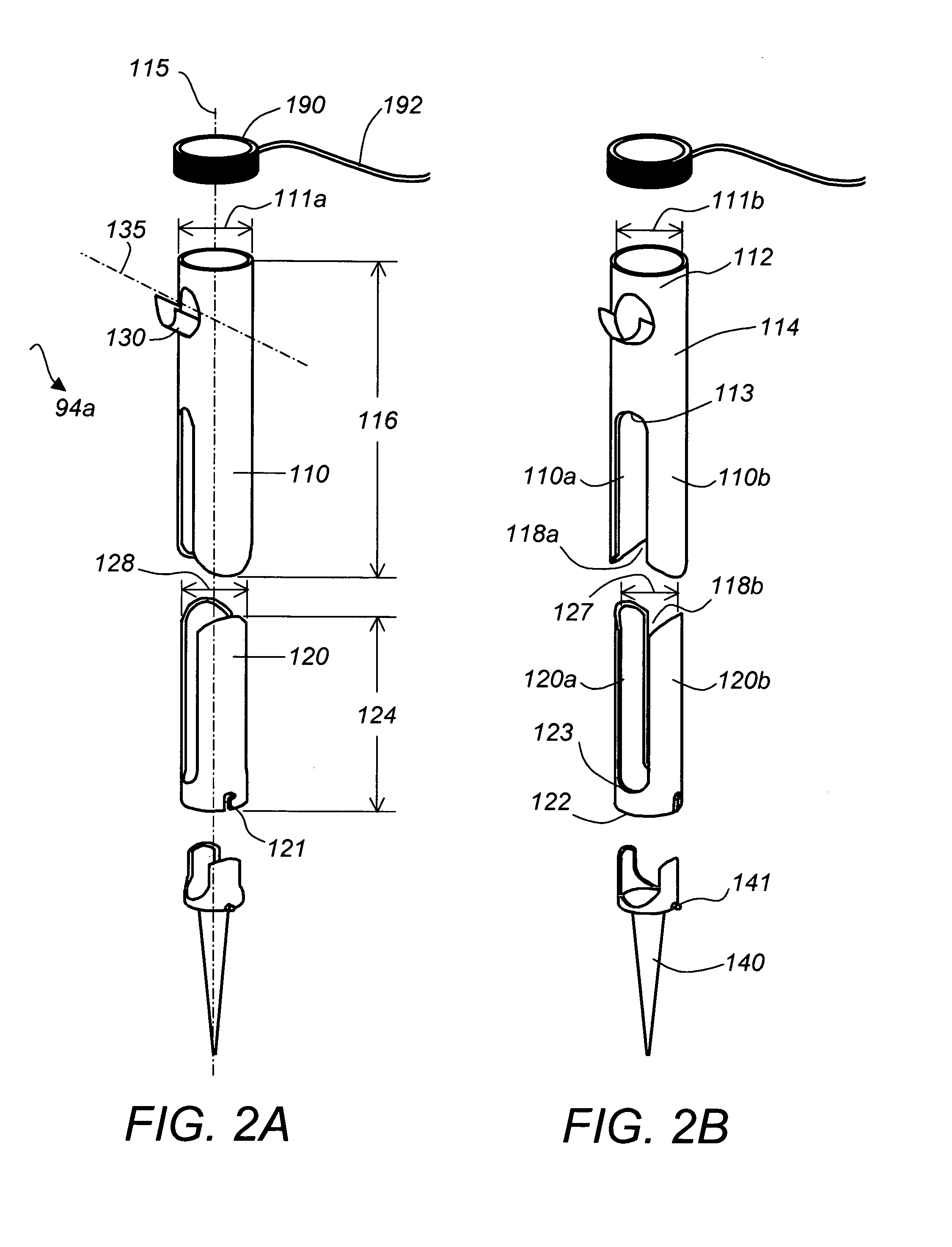
[0050] According to one embodiment of this invention, shown in FIG. 2A, and FIG. 2B, portal 94a includes an outer elongated cannula 110 and an inner elongated cannula 120. Inner cannula 120 slides within outer cannula 110 and is secured at different locations of the inner wall of the outer cannula 110, thereby forming a first working channel 115 with adjustable length. This is especially desirable for reaching locations within the patient's body corresponding to the outer locations 92a-92f, that are at different distances from the patient's skin 70. Outer cannula 110 has mill...


PUM


Abstract

A device for use as a portal in percutaneous minimally invasive surgery performed within a patient's body cavity includes a first elongated hollow tube having a length adjusted with a self-contained mechanism. The first elongated tube includes an inner hollow tube and an outer hollow tube and the inner tube is adapted to slide within the outer tube thereby providing the self-contained length adjusting mechanism. This length-adjustment feature is advantageous for percutaneous access surgery in any body cavity. Two or more elongated tubes with adjustable lengths can be placed into two or more adjacent body cavities, respectively. Paths are opened within the tissue areas between the two or more body cavities, and are used to transfer devices and tools between the adjacent body cavities. This system of two or more elongated tubes with adjustable lengths is particularly advantageous in percutaneous minimally invasive spinal surgeries, and provides the benefits of minimizing long incisions, recovery time and post-operative complications.

Description

CROSS REFERENCE TO RELATED CO-PENDING APPLICATIONS [0001] This application is a continuation in part of U.S. application Ser. No. 10 / 669,927 filed on Sep. 24th, 2003 and entitled APPARATUS AND METHOD FOR CONNECTING SPINAL VERTEBRAE the contents of which are expressly incorporated herein by reference. This application also claims the benefit of U.S. provisional application Ser. No. 60 / 518,580 filed on Nov. 8th, 2003 and entitled METHODS AND DEVICES FOR IMPROVING PERCUTANEOUS ACCESS the contents of which are expressly incorporated herein by reference.FIELD OF THE INVENTION [0002] The present invention relates to methods and devices for improving percutaneous access in minimally invasive surgeries, and more particularly to methods and devices that provide variable length access channels to locations deep within a patient's body, and allow the percutaneous transfer of connecting devices and instruments within one access channel or between two or more adjacent access channels placed deep...

Claims


Application Information

Patent Timeline
Patent Type & Authority Applications(United States)
IPC IPC(8): A61B17/00A61B17/34A61B17/88A61B19/00
CPCA61B17/00234A61B17/3421A61B17/7085A61B17/7083A61B2017/3443A61B2017/3445A61B19/54A61B90/39A61B17/7089A61B17/708A61B17/7088
Inventor CHIN, KINGSLEY RICHARD
Owner STRYKER EURO OPERATIONS HLDG LLC
Why Eureka
Try Eureka