Method for generating an X-ray image of an extremity of a patient with a scale of length

a technology of x-ray image and patient, which is applied in the direction of radiation beam directing means, patient positioning for diagnostics, instruments, etc., can solve the problem of real challenge in measuring the distance between the extremity of the patient and the x-ray detector

Inactive Publication Date: 2008-07-08
SIEMENS HEALTHCARE GMBH
View PDF10 Cites 12 Cited by
  • Summary
  • Abstract
  • Description
  • Claims
  • Application Information

AI Technical Summary

Problems solved by technology

While the distance from the X-ray source to the X-ray detector will generally be constant or can easily be made available by the control as an electronic data value, measurement of the distance from the extremity of the patient to the X-ray detector presents the real challenge.

Method used


Image

  • Method for generating an X-ray image of an extremity of a patient with a scale of length
  • Method for generating an X-ray image of an extremity of a patient with a scale of length
  • Method for generating an X-ray image of an extremity of a patient with a scale of length

Examples


Embodiment Construction

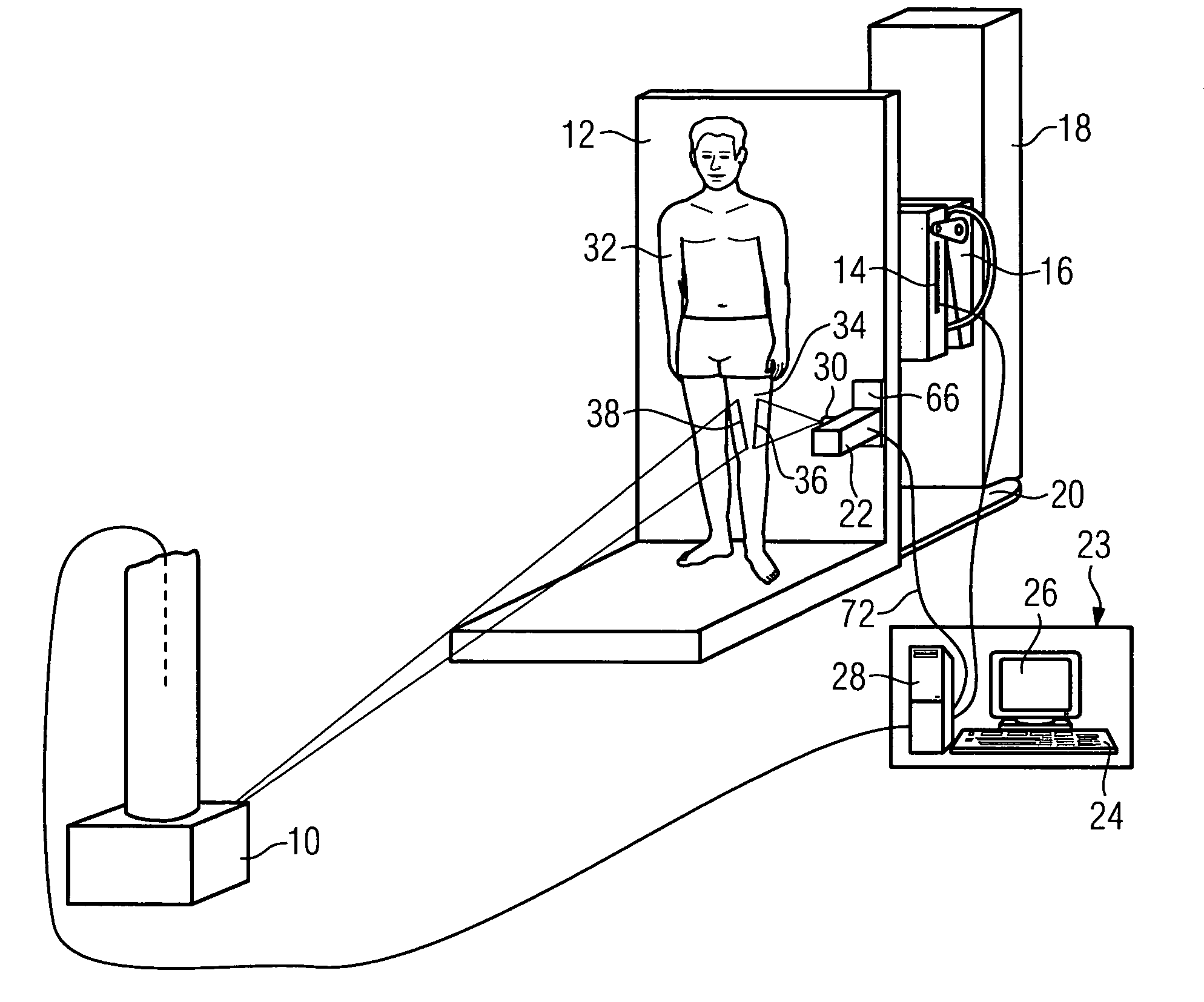
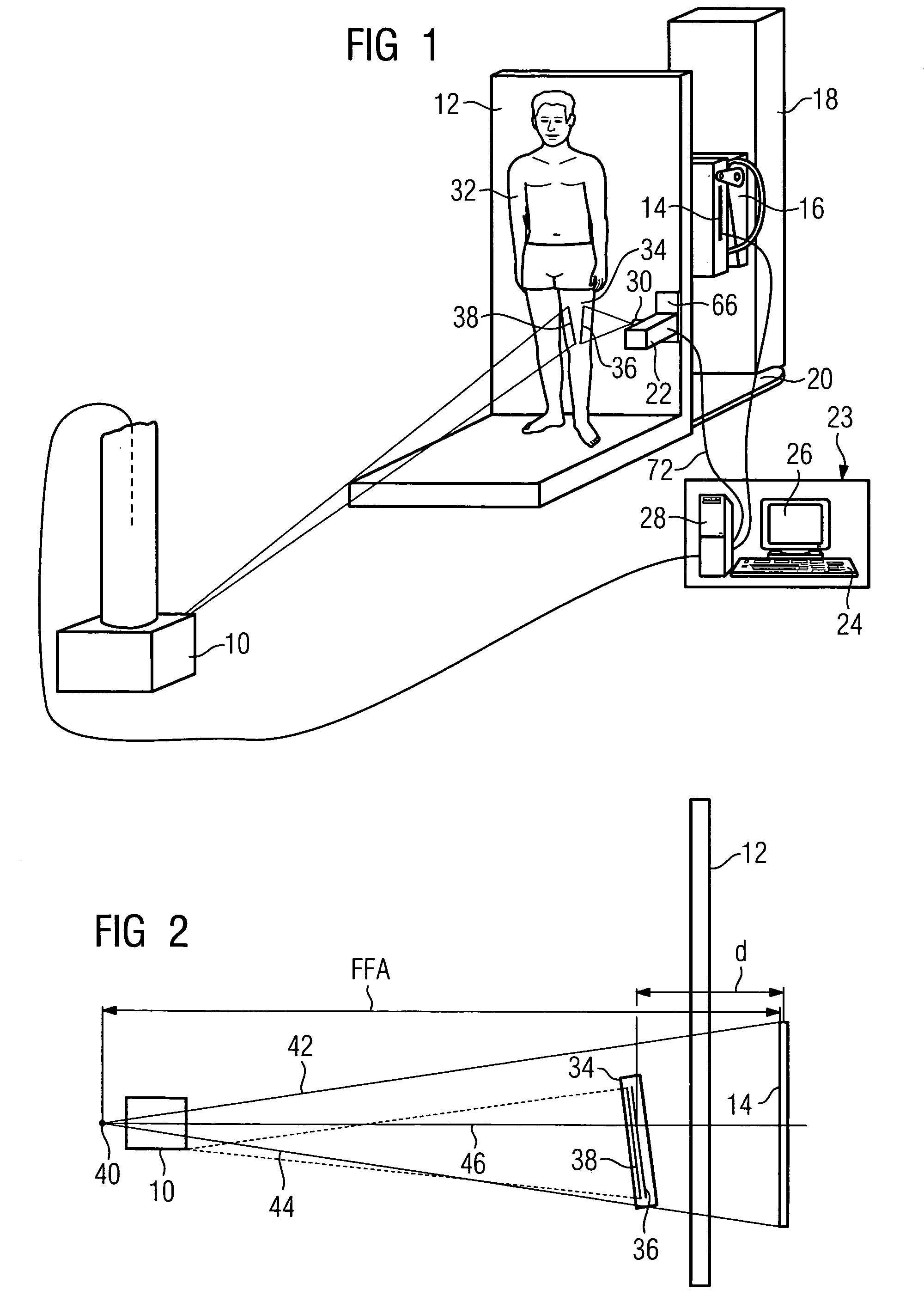
[0036]FIG. 1 shows an X-ray installation according to the invention. It comprises an X-ray source 10, a patient support 12 as a patient presentation support and an X-ray detector 14. The X-ray detector 14 is located in a holder 16 which is displaceable on an X-ray detector support 18. The patient support 12 stands not only simply in front of the X-ray detector support 18 but is arranged in a defined manner stationary in relation to this X-ray detector support. An adapter base 20 defines the distance from the patient support to the X-ray detector support and thus the distance from the patient support 12 to the X-ray detector 14.

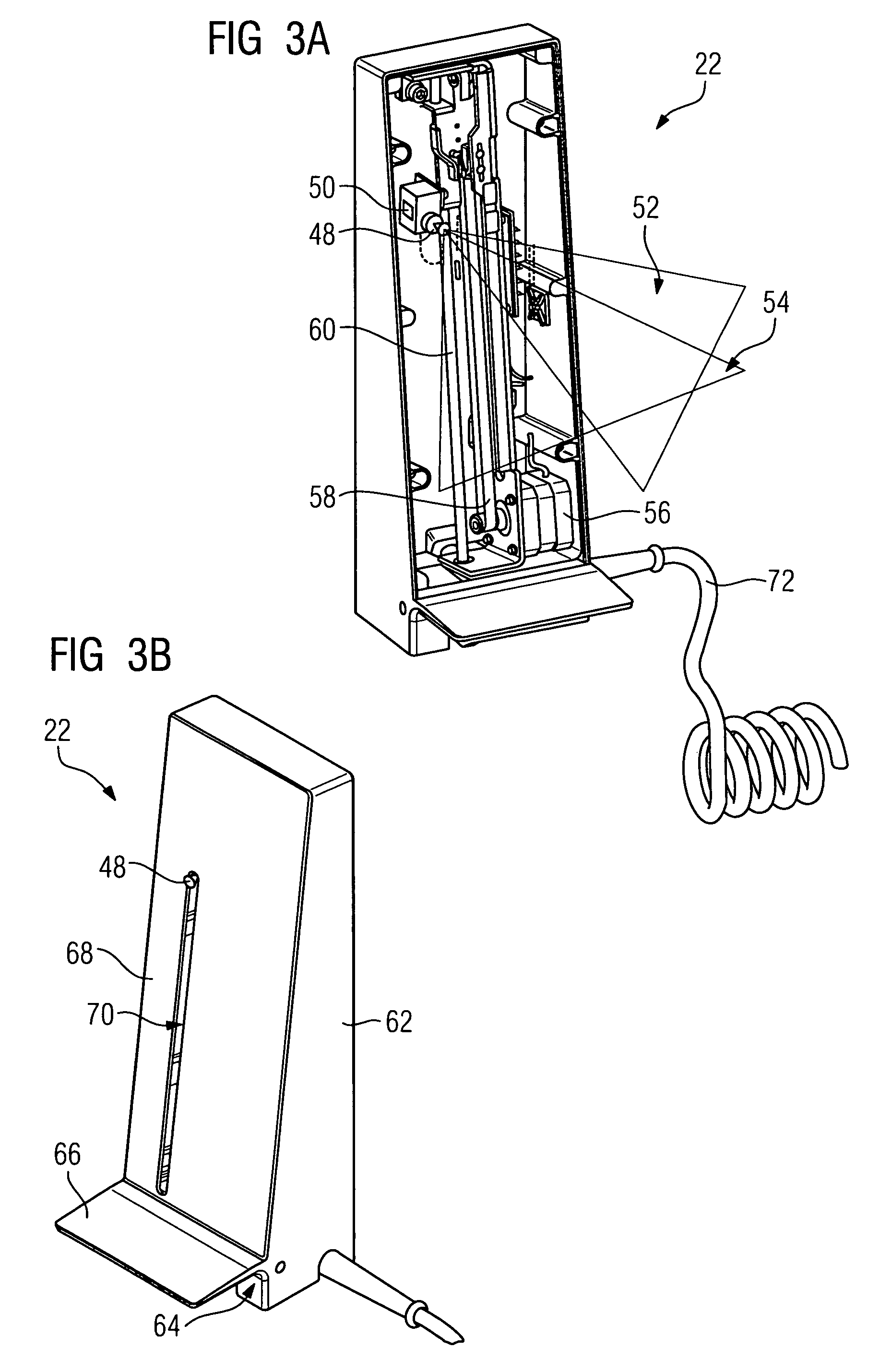
[0037]Standing in a stationary position relative to the patient support 123 is a light-source device 22, which will be described in greater detail later with reference to FIGS. 3A and 3B. The light-source device 22 can be vertically displaceable, but its distance from the X-ray detector 14 is defined and fixed.

[0038]Furthermore, part of the X-ray installation ...


PUM


Abstract

In an X-ray installation comprising an X-ray source, a patient support, an X-ray detector and an electronic evaluation system, a distance is determined from an extremity of a patient which is to be imaged to the X-ray detector and is made available as an electronic data value to the electronic evaluation system. If the distance from the X-ray source to the X-ray detector as an electronic data value is also known, then a scale can be calculated for the X-ray image from the two distances on the basis of the imaging laws for the generation of the X-ray image, which scale can be faded into the X-ray image or else electronically assigned to this image for an interrogation of coordinates.

Description

CROSS REFERENCE TO RELATED APPLICATIONS[0001]This application claims priority of German application No. 10 2005 046 764.4 DE filed Sep. 29, 2005, which is incorporated by reference herein in its entirety.FIELD OF INVENTION[0002]The invention relates to a method for generating an X-ray image of an extremity of a patient with a scale of length. It also relates to a light-source device which is used in such a method, as well as, in general, to an X-ray installation. Finally, the invention also relates to a patient support for an X-ray installation.BACKGROUND OF THE INVENTION[0003]Full information from an X-ray image can include absolute values of the lengths of extremities of a patient being known. Such absolute data can be indispensable, particularly in the planning of an operation.[0004]A lead ruler has been used until now to provide a scale of length. The lead ruler is arranged next to the patient or else fastened to him / her and is also reproduced on the X-ray image. In particular, ...

Claims


Application Information

Patent Timeline
Patent Type & Authority Patents(United States)
IPC IPC(8): H05G1/00G01D18/00
CPCA61B6/04A61B6/589A61B6/588A61B6/08
Inventor SCHMITT, THOMAS
Owner SIEMENS HEALTHCARE GMBH
Why Eureka
Try Eureka