
Medical closure clip system and method

a clip system and medical technology, applied in the field of medical devices, can solve the problems of time-consuming and laborious suturing, more difficult to approximate and close the same, and inability to join tissue structures, etc., to achieve the effect of reducing the formation of subcutaneous voids or pockets

Inactive Publication Date: 2008-08-12
3M INNOVATIVE PROPERTIES CO
View PDF200 Cites 110 Cited by
  • Summary
  • Abstract
  • Description
  • Claims
  • Application Information

AI Technical Summary

Benefits of technology

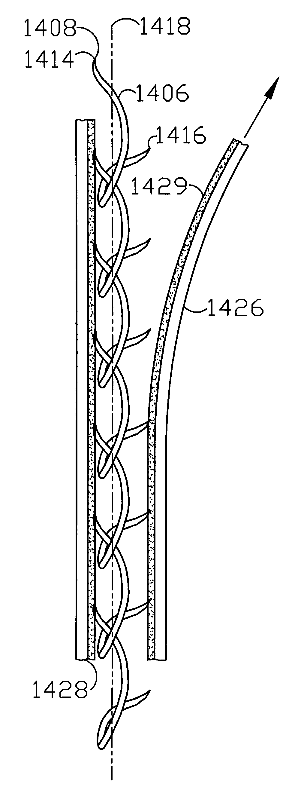
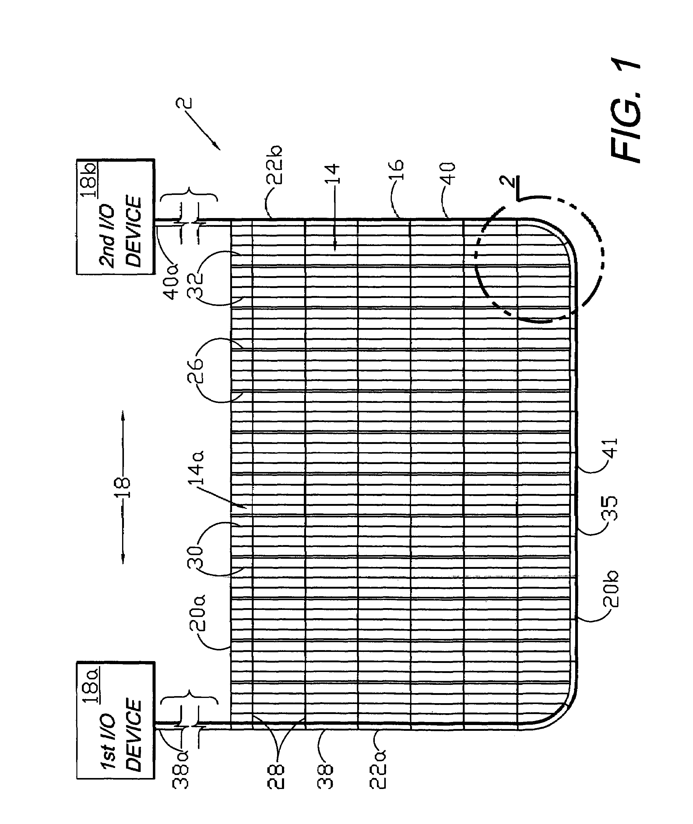
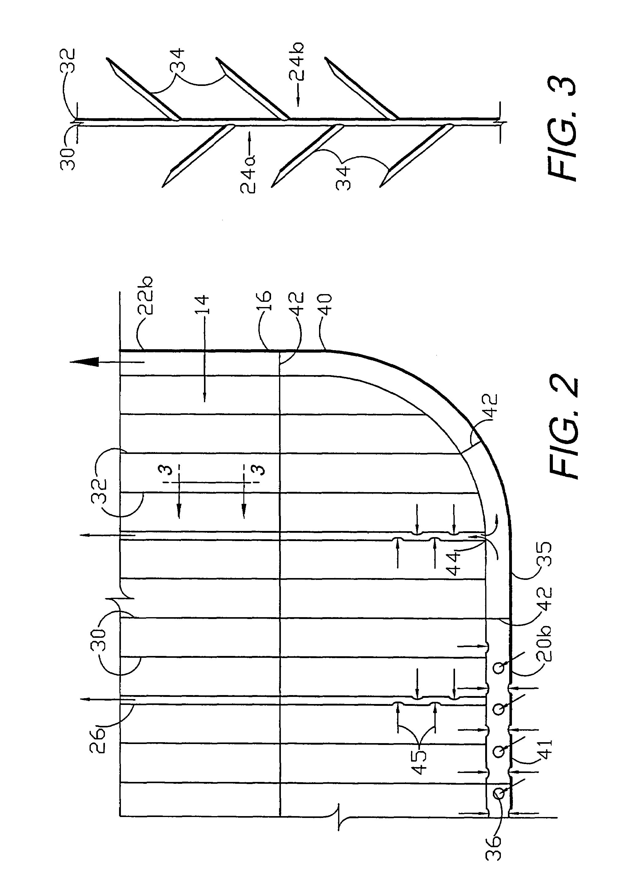
[0013]Alternative embodiment vertical risers are also disclosed, and can provide active fluid transfer utilizing the patient's body dynamics. Yet another alternative embodiment of the present invention utilizes the screen barbs for mechanical fixation in a separation for closure of same. Separation closure, irrigation and drainage methodologies are disclosed utilizing various combinations of closure screens, tubing, sutures, fluid transfer elements and gradient force sources. The closure screen of the present invention uses mechanical and other forces associated with screens and barbed strands for securing separated tissues together and for eliminating or reducing the formation of subcutaneous voids or pockets, which can potentially form hematoma and seroma effects. Further embodiments of the invention include assemblies of clips, which can comprise relatively rigid material, with flexible, elastic or collapsible connecting filaments forming composite material screens.

Problems solved by technology

They tend to occupy surgical teams and other resources for significant portions of many surgical procedures.
However, suturing can be time-consuming and tedious.
Moreover, the tissue structures to be joined may not be amenable to other closure techniques.
MIS often restricts access to the separated tissue structures, thus making it more difficult to approximate and close same.
Suturing in these extensive defects can be time-consuming and tedious.
Dead-space problems are more apt to occur in the subcutaneous closure.
However, deeper incisions may well require not only skin surface closure, but also time-consuming placement of multiple layers of sutures in the load-bearing planes.

Method used


Image

  • Medical closure clip system and method
  • Medical closure clip system and method
  • Medical closure clip system and method

Examples


Embodiment Construction

I. Introduction and Environment

[0077]As required, detailed embodiments of the present invention are disclosed herein; however, it is to be understood that the disclosed embodiments are merely exemplary of the invention, which may be embodied in various forms. Therefore, specific structural and functional details disclosed herein are not to be interpreted as limiting, but merely as a basis for the claims and as a representative basis for teaching one skilled in the art to variously employ the present invention in virtually any appropriately detailed structure.

[0078]Certain terminology will be used in the following description for convenience in reference only and will not be limiting. For example, the words “upwardly”, “downwardly”, “rightwardly” and “leftwardly” will refer to directions in the drawings to which reference is made. The words “inwardly” and “outwardly” will refer to directions toward and away from, respectively, the geometric center of the embodiment being described an...


PUM


Abstract

A medical closure screen device for a separation of first and second tissue portions is provided, which includes a mesh screen comprising tubular vertical risers, vertical strands with barbed filaments, and horizontal spacers connecting the risers and strands in a grid-like configuration. An optional perimeter member partly surrounds the screen and can comprise a perimeter tube fluidically coupled with the vertical risers to form a tubing assembly. Various input / output devices can optionally be connected to the perimeter tube ends for irrigating and / or draining the separation according to methodologies of the present invention. Separation closure, irrigation and drainage methodologies are disclosed utilizing various combinations of closure screens, tubing, sutures, fluid transfer elements and gradient force sources. The use of mechanical forces associated with barbed strands for repositionably securing separated tissues together is disclosed. The use of same for eliminating or reducing the formation of subcutaneous voids or pockets, which can potentially form hematoma and seroma effects, is also disclosed. The device can be fabricated and the method practiced with clips having various configurations.

Description

CROSS-REFERENCE TO RELATED APPLICATION[0001]This application is a continuation-in-part of U.S. patent application Ser. No. 10 / 224,852 filed on Aug. 21, 2002 currently pending in the United States Patent Office, and is incorporated herein by reference.BACKGROUND OF THE INVENTION[0002]1. Field of the Invention[0003]The present invention relates generally to medical (including dental, veterinary, etc.) closure and wound fluid management devices, and in particular to a screen closure member for closing tissue separations, such as incisions and wounds, which closure member is optionally bioabsorbable. The closure member can be assembled with different components chosen for their functional and material compatibility characteristics.[0004]2. Description of the Prior Art[0005]In the medical field, which is broadly defined to include dentistry, veterinary medicine, etc., cutaneous incisions are commonly performed in surgery to provide access to underlying tissue, organs, joints, skeletal st...

Claims


Application Information

Patent Timeline
Patent Type & Authority Patents(United States)
IPC IPC(8): A61B17/08A61D1/00A61B17/00A61B17/03A61B17/064A61M1/00
CPCA61B17/064A61B17/08A61B2217/005A61B2017/06176A61B2017/081A61B2017/00004
Inventor ZAMIEROWSKI, DAVID S.
Owner 3M INNOVATIVE PROPERTIES CO
Learn More