Apparatus and method for endoscopic encirclement of pulmonary veins for epicardial ablation

an endoscopic and pulmonary vein technology, applied in the field of applicatives and methods for endoscopic encirclement of pulmonary veins for epicardial ablation, can solve the problems of hampered encirclement of all four pulmonary veins with an epicardial ablation probe, difficult access, and hazardous dissection

Inactive Publication Date: 2009-10-06
MAQUET CARDIOVASCULAR LLC
View PDF197 Cites 46 Cited by
  • Summary
  • Abstract
  • Description
  • Claims
  • Application Information

AI Technical Summary

Problems solved by technology

In addition, encirclement of all four pulmonary veins with an epicardial ablation probe is hampered by two folds (known as reflections) of pericardium.
With the pericardial reflection between the superior vena cava and the right superior pulmonary vein, access is more difficult because this reflection forms the end of the transverse pericardial sinus and there is no direct way for the endoscopic subxiphoid cannula to approach this pericardial reflection.
Without direct access and good visualization of this pericardial reflection, dissection is hazardous because the superior vena cava, right superior pulmonary vein, and right main pulmonary artery are all in the vicinity of the reflection.
Grasping and forming a hole in the wrong structure, for example, the superior vena cava, would be disastrous.

Method used


Image

  • Apparatus and method for endoscopic encirclement of pulmonary veins for epicardial ablation
  • Apparatus and method for endoscopic encirclement of pulmonary veins for epicardial ablation
  • Apparatus and method for endoscopic encirclement of pulmonary veins for epicardial ablation

Examples


Embodiment Construction

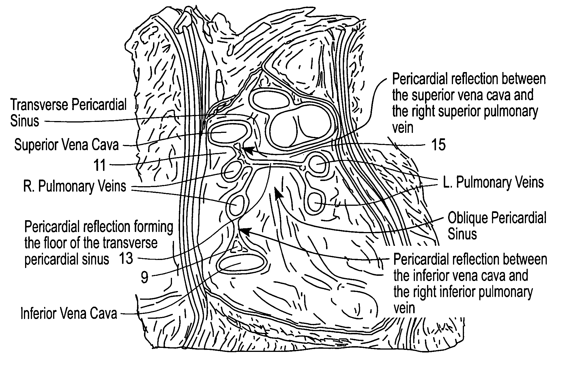
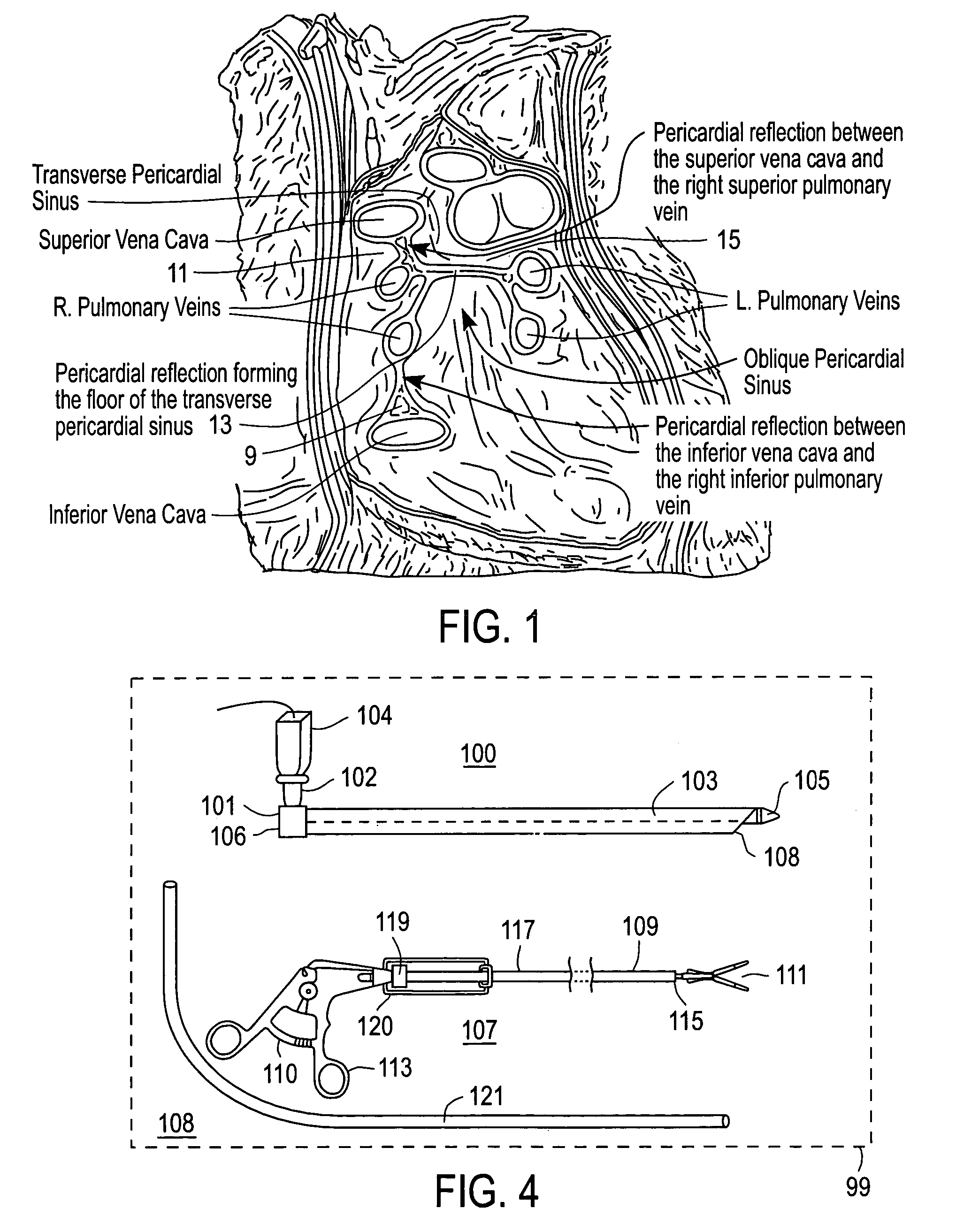
[0013]Referring now to the simplified anatomical illustration of FIG. 1, there is shown a view of the pericardial sac (with the heart absent), as viewed frontally. In this view, there is shown the reflection 9 disposed between the inferior vena cava and the right inferior pulmonary vein. Additionally, this view shows the reflection 11 disposed between the superior vena cava and the right superior pulmonary vein. Also, this view shows the reflection 13 that forms the base of the transverse pericardial sinus 15. The objective of the surgical procedure performed in accordance with an embodiment of the present invention is to encircle the right and left pulmonary veins with a tissue-ablation probe (or sheath through which the ablation probe may be positioned) in order to ablate atrial tissue along a path substantially encircling the ostia of these veins. This is accomplished with diminished risk of penetration of the veins and arteries in the vicinity and with minimal damage or trauma t...


PUM


Abstract

Surgical instruments and procedures promote placement of a tissue-ablating probe surrounding the left and right pulmonary veins within the intrapericardial space of a patient's heart via access through a subxiphoid or subcostal entry incision. An opening is formed in the pericardium near the apex region of the heart to facilitate formation of openings through three pericardial reflections by which a tissue-ablating probe is positioned around the pulmonary veins.

Description

RELATED APPLICATIONS[0001]This application is a continuation-in-part of application Ser. No. 10 / 369,980, entitled “Subxiphoid Procedures And Apparatus For Placement Of Cardiac Defibrillator And Pacer”, filed on Feb. 18, 2003 now U.S. Pat. No. 7,288,096 by Albert K. Chin, which is a continuation-in-part application of application Ser. No. 10 / 347,212, entitled “Apparatus And Methods For Endoscopic Surgical Procedures”, filed on Jan. 17, 2003 now abandoned by Albert K. Chin, and which is also a continuation-in-part of application Ser. No. 10 / 346,663 entitled “Endoscopic Subxiphoid Surgical Procedures”, filed on Jan. 17, 2003 now U.S. Pat. No. 7,264,587 by Albert K. Chin, et al., which is a continuation-in-part of pending application Ser. No. 10 / 140,309 entitled “Methods And Apparatus For Endoscopic Cardiac Surgery”, filed on May 6, 2002 by Albert K. Chin, et al., which is a continuation of pending application Ser. No. 09 / 635,721 entitled “Apparatus For Endoscopic Access”, filed on Aug....

Claims


Application Information

Patent Timeline
Patent Type & Authority Patents(United States)
IPC IPC(8): A61B17/32A61B18/14A61BA61B1/01A61B17/00A61B17/06A61B17/30A61B17/34A61B19/00A61N1/05
CPCA61B17/00008A61B19/201A61N1/0587A61B1/00154A61B17/3468A61B1/01A61B17/3478A61B18/1445A61B2017/00243A61B2017/061A61B2017/306A61B2017/320044A61B2018/00291A61B2019/306A61N2001/0578A61B90/11A61B2090/036
Inventor CHIN, ALBERT K.
Owner MAQUET CARDIOVASCULAR LLC
Why Eureka
Try Eureka