Method of calibration of digital X-ray apparatus and its embodiments

a digital x-ray and calibration method technology, applied in the field of medical xray engineering, can solve the problems of significant deterioration of an image being reconstructed, calibration of shading, failure of the preliminary calibration of the detector, etc., and achieve the improvement of mechanical precision tolerance, the effect of improving the calibration accuracy of the digital x-ray apparatus

Inactive Publication Date: 2010-01-19
ZAO IMPULSE
View PDF15 Cites 3 Cited by
  • Summary
  • Abstract
  • Description
  • Claims
  • Application Information

AI Technical Summary

Benefits of technology

[0012]The objective of the invention is to create higher precision calibration method for X-ray apparatuses. The technical result of the invention according to the first and the second variants is the improvement of digital X-ray apparatus calibration precision due to formation of the calibration data set for the stand regular component of mechanical instability with due account of the scale calibration data, and the increasing of mechanical precision tolerance of the X-ray apparatus stand rotation, in case of using the methods under application. In the second variant, the determination precision of calibration data for the mechanical instability regular component of X-ray apparatus is higher than the precision in the first one due to the use, during calibration, of at least two balls of different diameters. The application of calibration data to X-ray images reduces the image noise that in its turn improves the resolution of low-contrast and high-contrast roentgenograms of X-ray apparatus, owing to what, in the medical X-ray diagnostics, for example, it is possible to detect malignant neoplasms at an earlier stage.

Problems solved by technology

So for example, the changes in digital X-ray apparatus stand rotation path may result in failures of the preliminary calibrations of the detector, such as calibration of shading, etc.
For the computed tomography apparatuses, besides the failure of the detector preliminary calibrations, even smallest changes in the path of the tomographic scanning may result in considerable deterioration of an image being reconstructed.
The obstacles to achieve the said below technical result, when using the known method, include, besides the complexity and the expensiveness of the given method application, possible obstructing of the optical stereo camera field of view (the marker becomes invisible for the camera during the apparatus rotation when the patient's table stands between the camera and the marker or when a medical staff person intrudes into the camera field of view, etc.) Thus, a part of diagnostic data will be obtained without corrections for mechanical instability of the apparatus, which circumstance leads to deterioration of the X-ray images being obtained.
The obstacles to achieve the said below technical result, when using the known method, include, besides the complexity and the expensiveness of the given method application, the fact that the ultrasonic radiation characteristics depend on the temperature of the medium through which it propagates.
This leads to necessity of complex calculations and correction of the information received by the ultrasonic data receivers, because the temperatures of X-ray tube, X-ray sensor and the ambient air are different.
The obstacles to achieve the said below technical result, when using the known method include the fact, that the test objects of this method are made of X-ray contrast material, of a metal as a rule.
Presence of such objects during each exposure causes the formation of artifacts on the image being reconstructed; that leads to inaccuracies when X-ray tomographic examining.
In the method, the leading role belongs to X-direction and Y-direction calibration data of X-ray apparatus stand mechanical instability, the obstacles to achieve the said below technical result using the known method include the fact, that in the known method the regular component of digital X-ray apparatus stand, Z-direction mechanical instability (scale calibration data) is not determined.

Method used


Image

  • Method of calibration of digital X-ray apparatus and its embodiments
  • Method of calibration of digital X-ray apparatus and its embodiments
  • Method of calibration of digital X-ray apparatus and its embodiments

Examples


Embodiment Construction

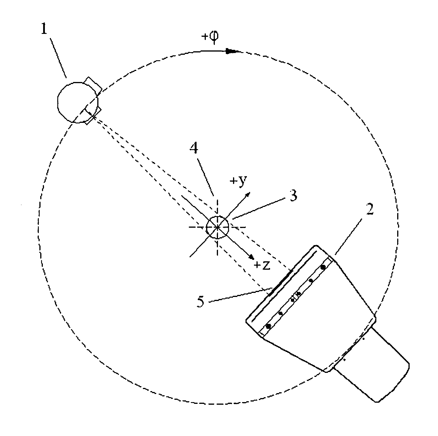
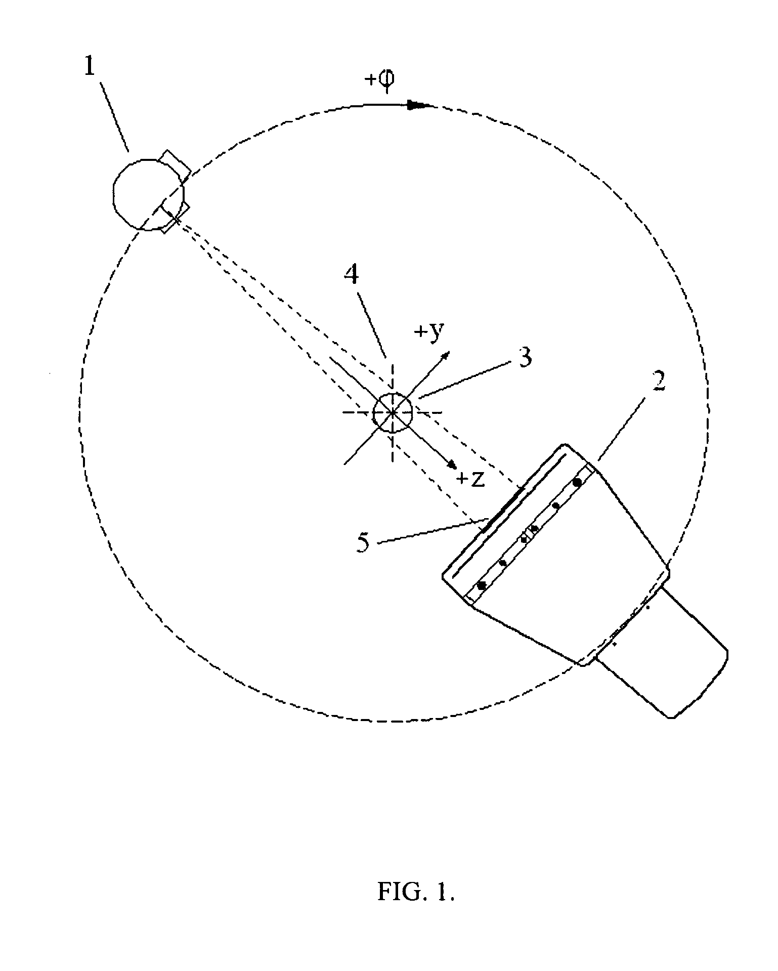
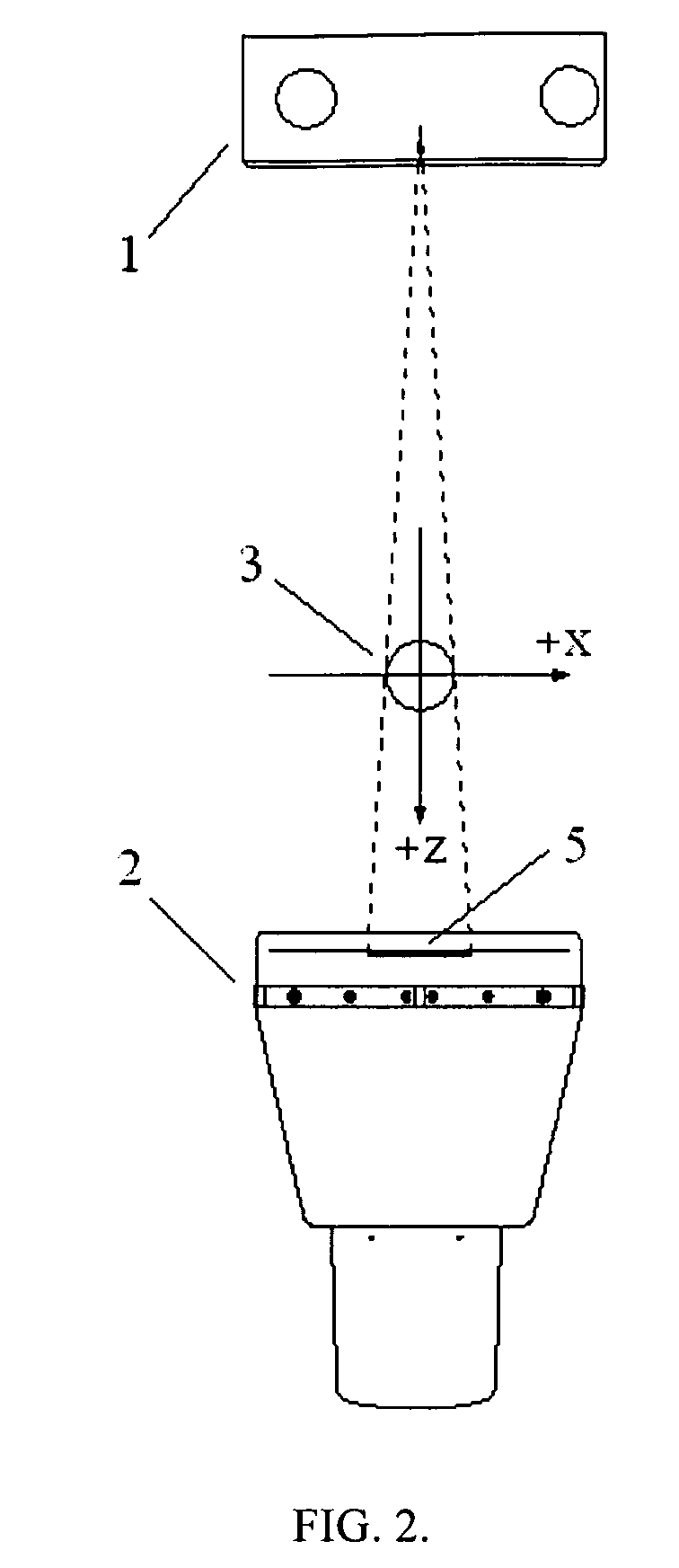
[0025]The method according to the first variant of the invention is to be implemented by means of device (FIG. 1, 2). The FIG. 1, 2 schematically shows the layout of digital X-ray apparatus, where:

[0026]1—X-ray tube,

[0027]2—detector,

[0028]3—ball of X-ray contrast material,

[0029]4—centre of the scanning path,

[0030]5—projection of the ball of X-ray contrast material.

[0031]In the second variant at least two balls made of X-ray contrast material, for example of steel, of different diameters, placed in the scanning field, excluding its centre are used as a calibration object.

[0032]The preliminary calibration of digital X-ray apparatus stand which includes X-ray tube 1 and detector 2 according to the first variant of the invention is to be implemented the following way. The ball 3 of X-ray contrast material, for example from steel, is placed in the scanning field excluding its centre 4 (the precision of the ball fabrication shall be higher than the spatial resolution of the digital X-ray ...


PUM


Abstract

The inventions (variants) are intended for improvement of calibration precision of digital X-ray apparatuses: computed tomographs, as well as digital X-ray apparatuses equipped with C-arm and U-arm type stands, which method is based on calculation of mechanical instability of the stand. Calibration method for X-ray apparatus, in which one or two X-ray contrast balls of different diameters are placed in the scanning field, excluding its centre, a series of images of the scanning field with different angles of the stand rotation are obtained, the X-direction and Y-direction regular components of mechanical instability of the X-ray apparatus stand are determined using the coordinates of projection centre of the ball, X-direction and Y-direction calibration modifications set for mechanical instability of the stand are formed, which modifications are used for correction of the images, according to the invention the additional regular component of mechanical instability of digital X-ray stand is determined using the scales of projections, and the calibration data set for mechanical instability of the stand, which includes the scale calibration modifications, is formed.

Description

RELATED APPLICATIONS[0001]This application is a Continuation of PCT application serial number PCT / RU2008 / 000308, filed on May 19, 2008, which claims priority to Russian Patent Application No. 2007140023, filed on Oct. 31, 2007, both of which are incorporated herein by reference in their entirety.FIELD OF THE INVENTION[0002]The group of inventions belongs to medical X-ray engineering, namely to the check and measurement instruments for technical characteristics of digital X-ray apparatuses: computed tomographs as well as digital X-ray apparatuses equipped with C-arm and U-arm type stands.BACKGROUND OF THE INVENTION[0003]The principal purpose of digital X-ray apparatus calibration is a production of highest quality images of the object under examination (patient) in course of radiological exploration. The quality of the explorative images is highly influenced by mechanical instability of the X-ray computed tomograph stand (also known as tomograph gantry) and of the C-arm and U-arm sta...

Claims


Application Information

Patent Timeline
Patent Type & Authority Patents(United States)
IPC IPC(8): G01D18/00
CPCA61B6/583A61B6/032
Inventor REBONI, VOLDEMAR O.JURENJA, VITALY V.SCHIRIY, ANDREI O.
Owner ZAO IMPULSE
Why Eureka
Try Eureka