Biopsy devices and methods

a biopsies and clip technology, applied in the field of breast implantation clips, can solve the problems of not always certain that the lesions are one and the same, the clip used is generally prone to pinching a minute amount of breast tissue, and the clip is not always easy to hold onto the tissu

Inactive Publication Date: 2010-03-02
SELIS JAMES E
View PDF36 Cites 49 Cited by
  • Summary
  • Abstract
  • Description
  • Claims
  • Application Information

AI Technical Summary

Benefits of technology

[0013]In one aspect, the present invention meets the above needs by providing an improved clip for mammography analysis, comprising a first portion that is straight, arcuate or a combination thereof; and at least a second portion that is straight, arcuate or a combination

Problems solved by technology

In addition, if something is seen on both mammography and ultrasound, it is not always certain that the lesions are one and the same.
The clip that is employed is generally prone to pinching a minute amount of breast tissue.
Sometimes, the clip may fail to hold onto the tissue or the clip may migrate to a different undesired location.
Recent indications are that U.S. Surgical may no longer manufacture this clip, thus creating a potential supply issue for existing users.
The pellets, however, potentially result in undesired migration of particles.
Although there is current production of a hand-held Mammotome® device, for the purpose of ultrasound

Method used


Image

  • Biopsy devices and methods
  • Biopsy devices and methods
  • Biopsy devices and methods

Examples


Embodiment Construction

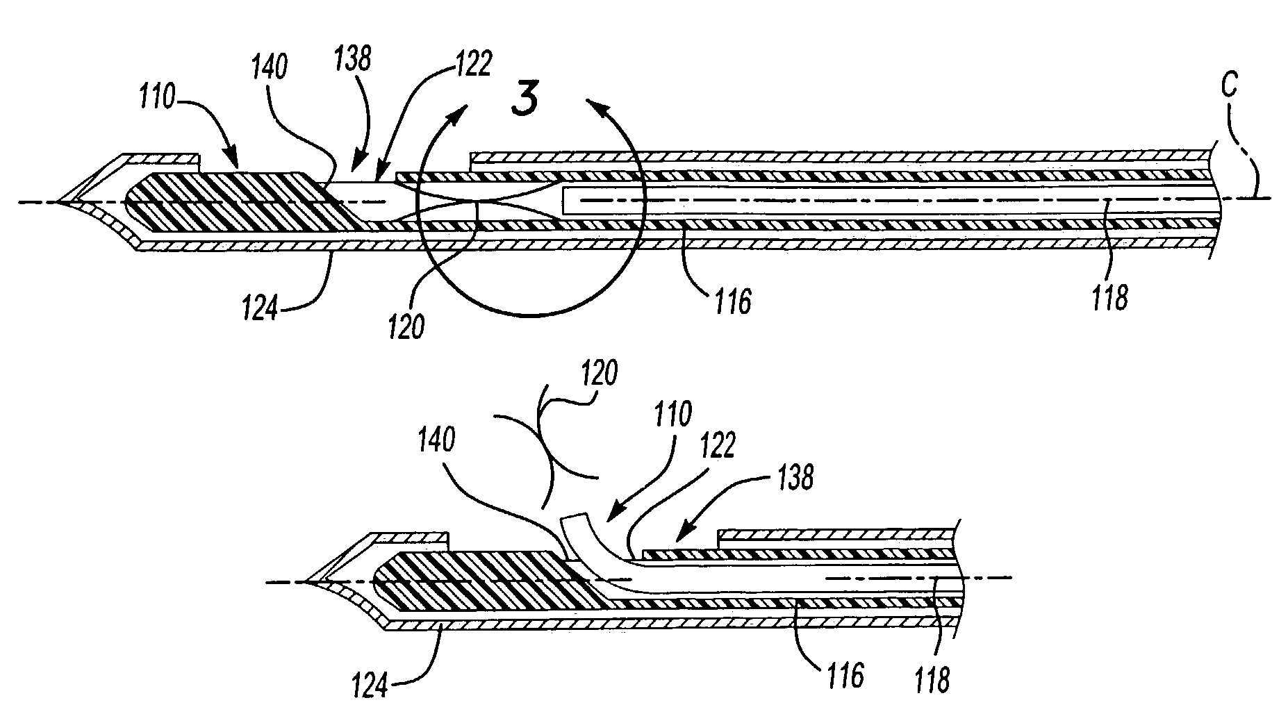
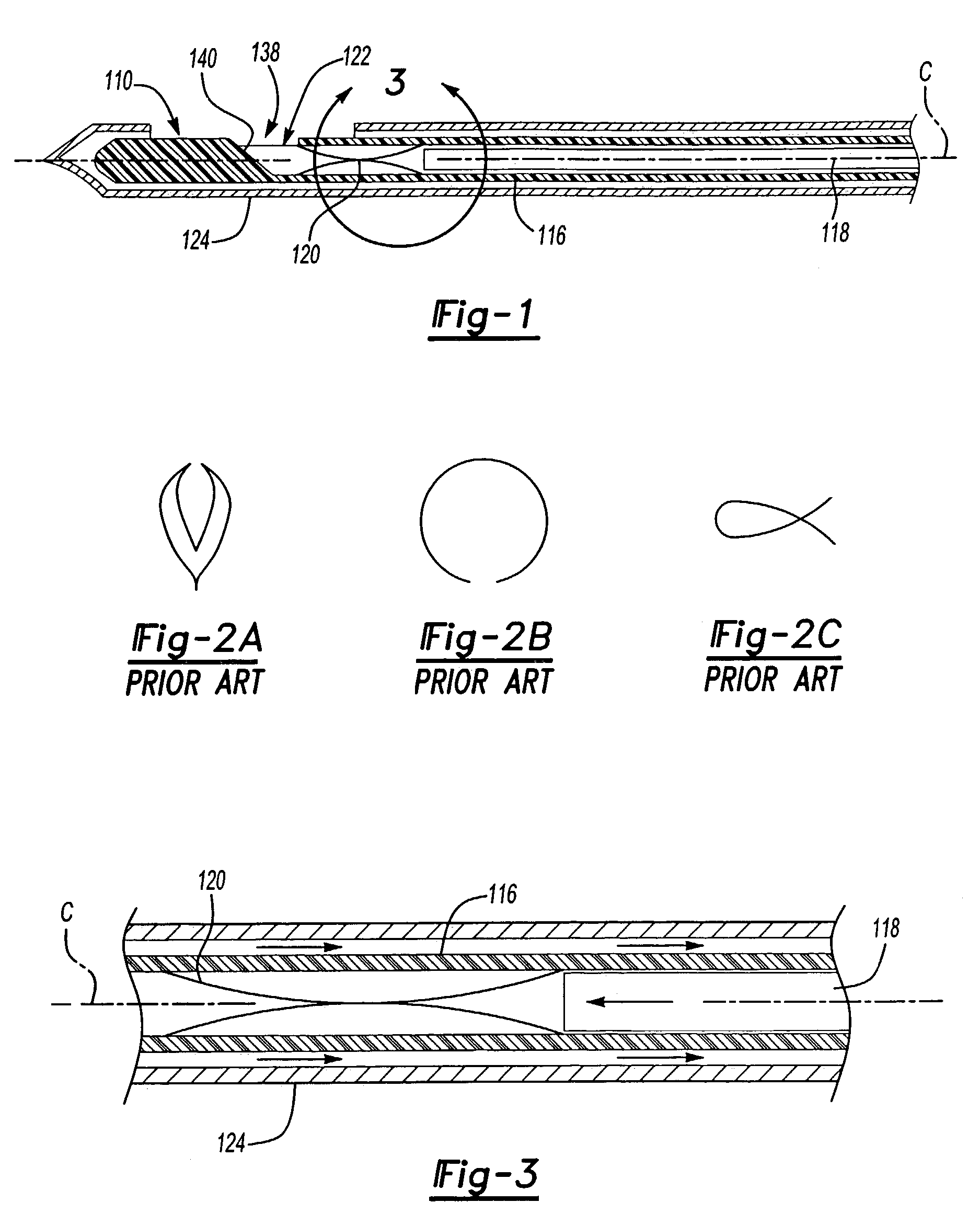
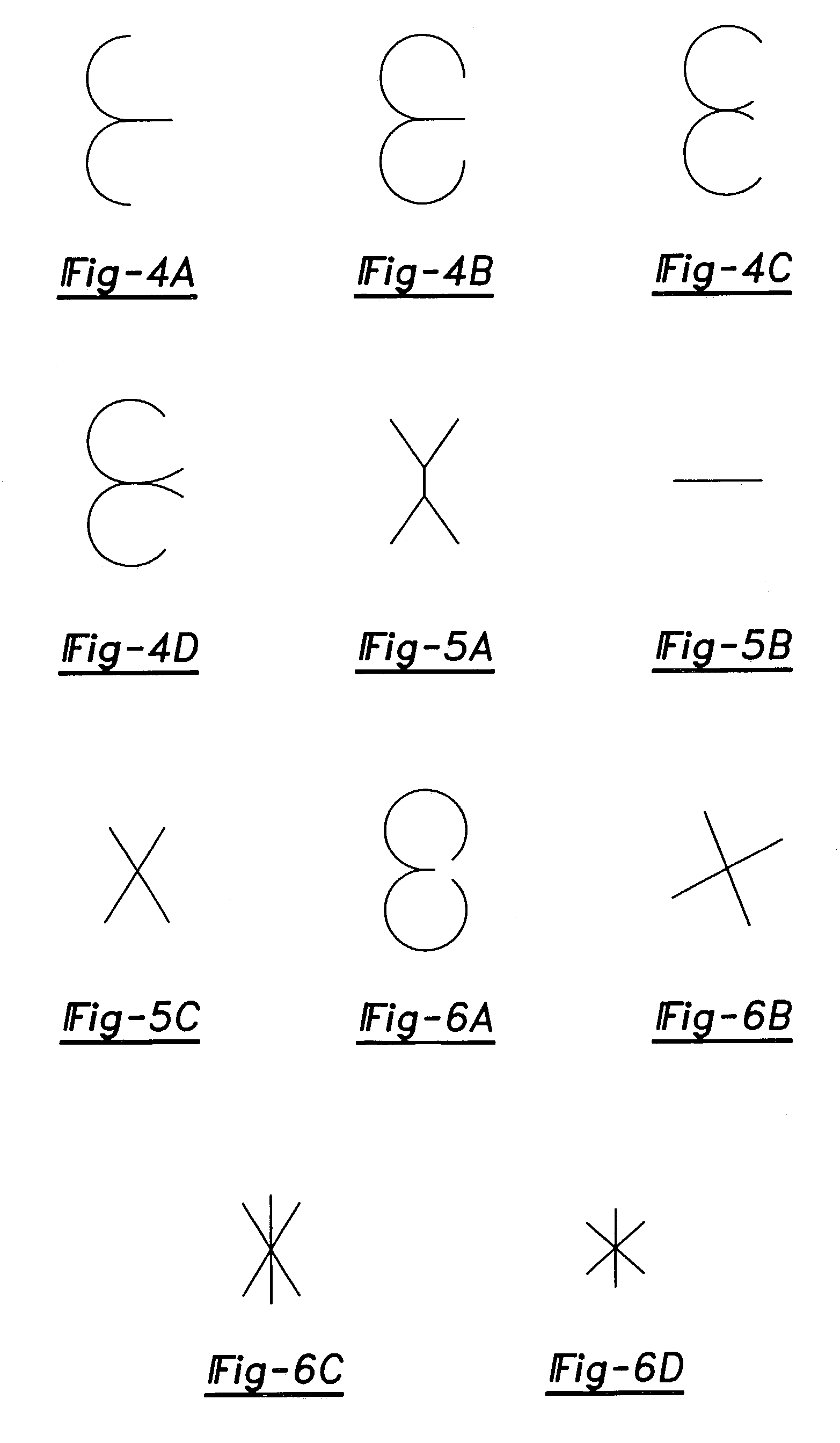
[0035]The present invention is directed to improved devices and methods for medical diagnosis and treatment, and particularly devices that are employed for mammographic analysis, such as for the detection and treatment of cancerous or other abnormal growths. The present invention contemplates an improved clip, such as for use as a marker, a delivery device for deploying the clip, a cyst aspiration device, combinations thereof and methods of using the same.

[0036]As will be seen from the description that follows, the various inventive features are not confined to a single application, but rather are capable of numerous variations. Accordingly, though described in a certain context, as will be apparent, features may be interchangeable among embodiments. For sake of brevity, while still providing ample instruction to the skilled artisan, the features herein are described without limitation in embodiments featuring the employment of a clip delivery device 110 (110′) by itself, with a bio...


PUM


Abstract

An improved system for mammography analysis and methods of using the same. In one aspect, a clip comprises a first portion and at least one additional second portion connected to the first portion, the first and second portions adapted for elastic deformation for engaging tissue. In another aspect, an improved actuator for performing a breast biopsy is used to deploy a clip. In yet another aspect of the invention, there is contemplated a system for marking an evacuated breast cyst.

Description

CLAIM OF BENEFIT OF FILING DATE[0001]The present application claims the benefit of the filing date of U.S. Provisional Application Ser. No. 60 / 400,113 (filed Aug. 1, 2002), the contents of which are hereby incorporated by reference in their entirety.TECHNICAL FIELD[0002]The present invention relates to devices and methods for performing biopsies, and more particularly to breast implantation clips for use as markers in mammography.BACKGROUND[0003]Percutaneous biopsy of the breast is a well-accepted alternative to open surgical biopsy with needle localization for those lesions seen by mammography or ultrasound but not able to be felt by the surgeon. When percutaneous biopsy is performed, it is frequently necessary to place a metal clip at the site of biopsy. This is done for several reasons. For example, the lesion biopsied might be partially or entirely removed. If the lesion is proven to be malignant, it is necessary to subsequently do a wide excisional biopsy after needle localizat...

Claims


Application Information

Patent Timeline
IPC IPC(8): A61B17/08A61B19/00
CPCA61B19/54A61B2019/5408A61B90/39A61B2090/3908
Inventor SELIS, JAMES E.
Owner SELIS JAMES E
Why Eureka
Try Eureka