

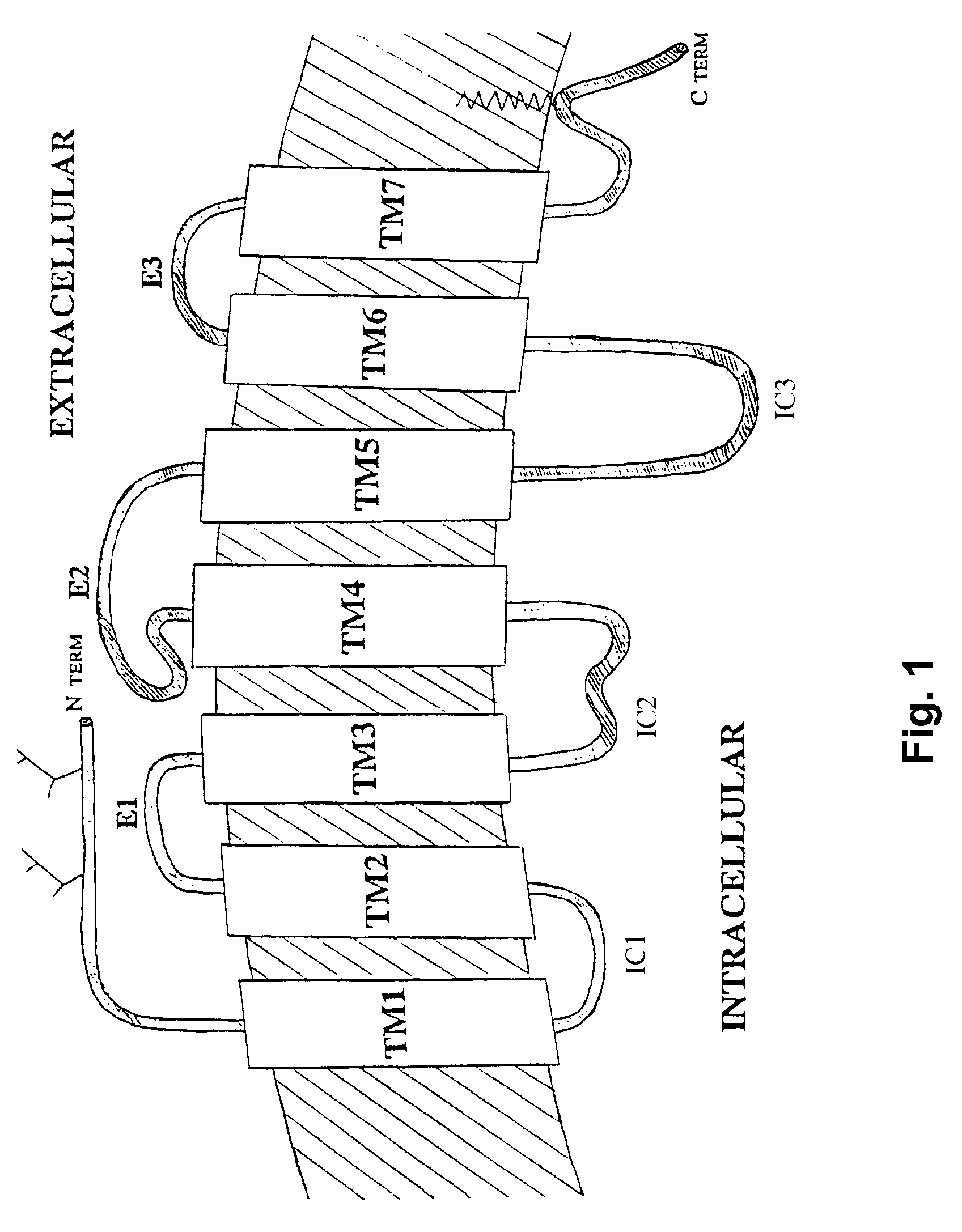
Method for obtaining G protein-coupled receptor (GPCR) diffraction-quality crystals employing a monoclonal antibody that binds to the third intracellular loop (IL3)
a technology of g protein-coupled receptor and monoclonal antibody, which is applied in the field of obtaining g protein-coupled receptor (gpcr) diffraction-quality crystals, can solve the problems of short extracellular and intracellular domains, poor structure,
- Summary
- Abstract
- Description
- Claims
- Application Information
AI Technical Summary
Problems solved by technology
Method used
Image

Examples
example 1
Antigen Production
[0092]Antigen was prepared by reconstituting purified, functional β2AR at high density into phospholipid vesicles (1 mg receptor per mg of phospholipids) using the following method.
[0093]β2AR having an amino terminal Flag epitope tag and a carboxyl hexahistidine tag was expressed in Sf9 insects cells and purified by sequential M1 antibody and alprenolol affinity chromatography as previously described (Kobilka Anal Biochem 1995 231, 269-71). Purified β2AR was immobilized on a Ni column and equilibrated with 10 column volumes of a mixture of 5 mg / ml DOPC (Avanti Polar Lipids) and 0.5 mg / ml Lipid A (Sigma) in 1% (w / v) octylglucoside (Anatrace), 100 mM NaCl, 20 mM Hepes pH 7.5 and 1 μM carazolol (a β2AR antagonist). The β2AR was then eluted in the same buffer containing 200 mM imidazole. The concentration of the eluted β2AR was adjusted to 5 mg / ml. This usually involved diluting the protein with the same buffer, but occasionally required concentrating the protein up to...
example 2
Antibody Production
[0095]Monoclonal antibodies (MABs) were generated using a conventional fusion protocol, as follows:
Myeloma Cell Lines
[0096]The P3x63Ag8.653 myeloma cell line (ATCC # CRL-1580; Lot No. 1131010) was used for the fusion experiment performed with splenocytes from immunized mice.
Cell Culture
[0097]Myeloma cells were propagated in Dulbecco's Modified Eagle's Medium (DMEM-high glucose) supplemented with 10% Fetal Bovine Serum (FBS), 0.15 g / L oxaloacetate, 0.05 g / L pyruvate, 0.0082 g / L bovine insulin (OPI supplement), 10% NCTC109 medium and 4 mM L-glutamine. This formulation is referred to the SDMEM formulation. Newly formed hybridoma were selected in SDMEM supplemented with HAT mixture (1.0×10⁻⁴ M Hypoxanthine, 4.0×10⁻⁶ M Aminopterin, 1.6×10⁻⁵ M thymidine), 5% hybridoma cloning factor, gentamicin (50 μg / ml) and β-mercaptoethanol at 0.055 mM. This formulation is referred as SDMEM-HAT. After selection, the newly formed hybridomas were propagated in SDMEM supplemented with 2...
example 3
Antibody Screening
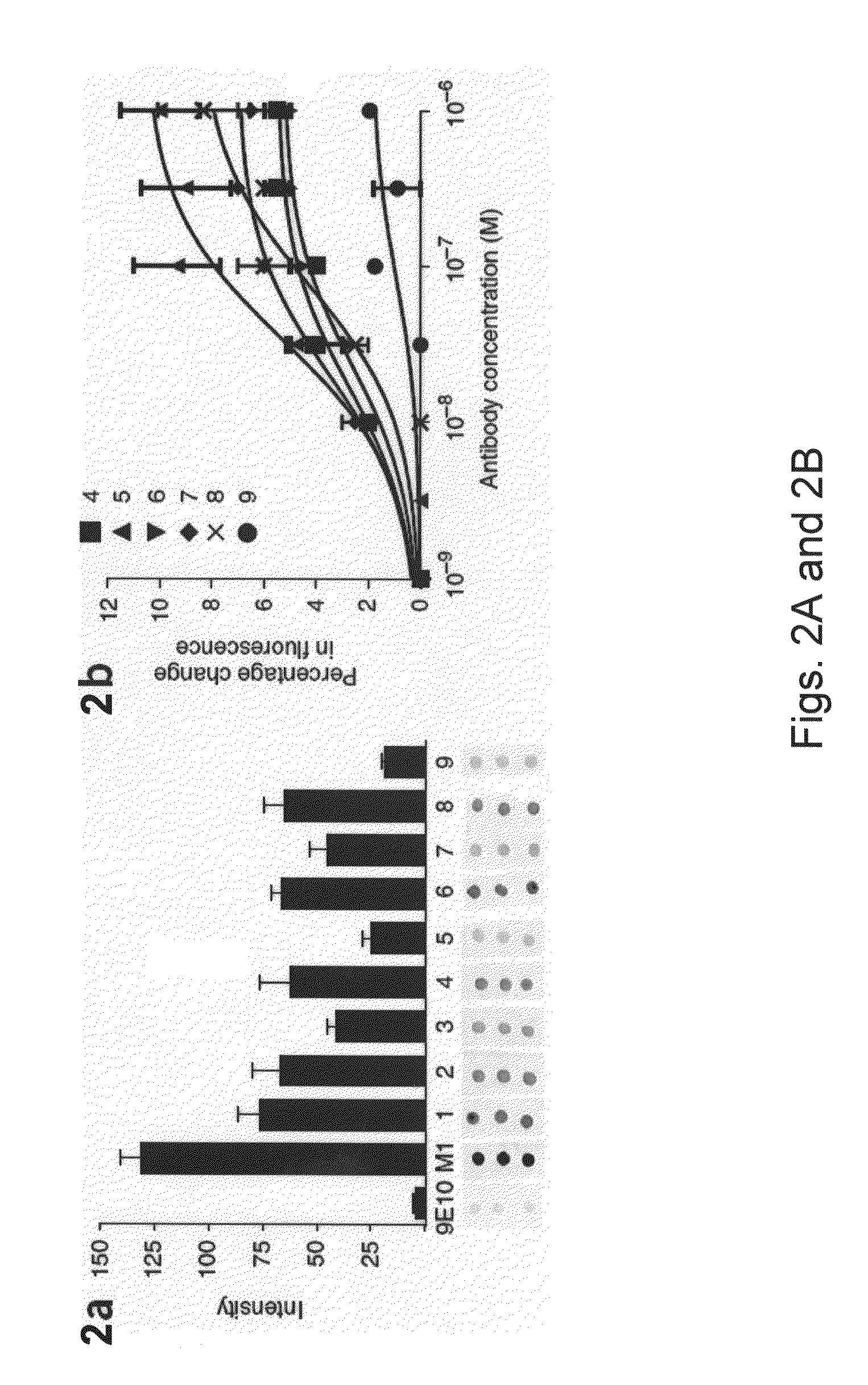
[0100]Antibodies were screened for binding to a three dimensional epitope on the three-dimensional surface on the IC3 of the native β2AR, rather than a flexible linear epitope. Nine MABs were screened, along with positive (M1 antibody) and negative (9E10) controls, for binding to β2AR denatured with sodium dodecyl sulfate (SDS) and spotted on nitrocellulose (FIG. 2A). Antibody 5 and antibody 9 showed the weakest binding to denatured protein, even though they showed immunostaining comparable to M1 on binding to native receptor in fixed cells (see FIG. 4). The reduced binding of antibody 5 and antibody 9 to SDS denatured β2AR showed that these antibodies bind to a three-dimensional epitope.
[0101]Antibodies 2, 5, 6, 7 and 9 all reacted with intracellular epitopes. To select for antibodies that may interact with IC3, we examined the effect of antibody binding on the fluorescence of β2AR labeled at C265 (at the cytoplasmic end of TM6) with tetramethylrhodamine. Tetramet...
PUM
| Property | Measurement | Unit |
|---|---|---|
| pH | aaaaa | aaaaa |
| pH | aaaaa | aaaaa |
| concentration | aaaaa | aaaaa |
Abstract
Description
Claims
Application Information