

Antibodies to non-shed Muc1 and Muc16, and uses thereof
a technology of antibodies and muc16, which is applied in the field of antibodies directed to plasma membrane epitopes, can solve the problems of limited antibody production methods and cannot be reliably detected, and achieve the effect of improving the properties of detection, monitoring and treatmen
- Summary
- Abstract
- Description
- Claims
- Application Information
AI Technical Summary
Benefits of technology
Problems solved by technology
Method used
Image

Examples
example 1
Generation of Monoclonal Antibodies to the Extracellular Cell-Associated Domains of Muc1 and Muc16
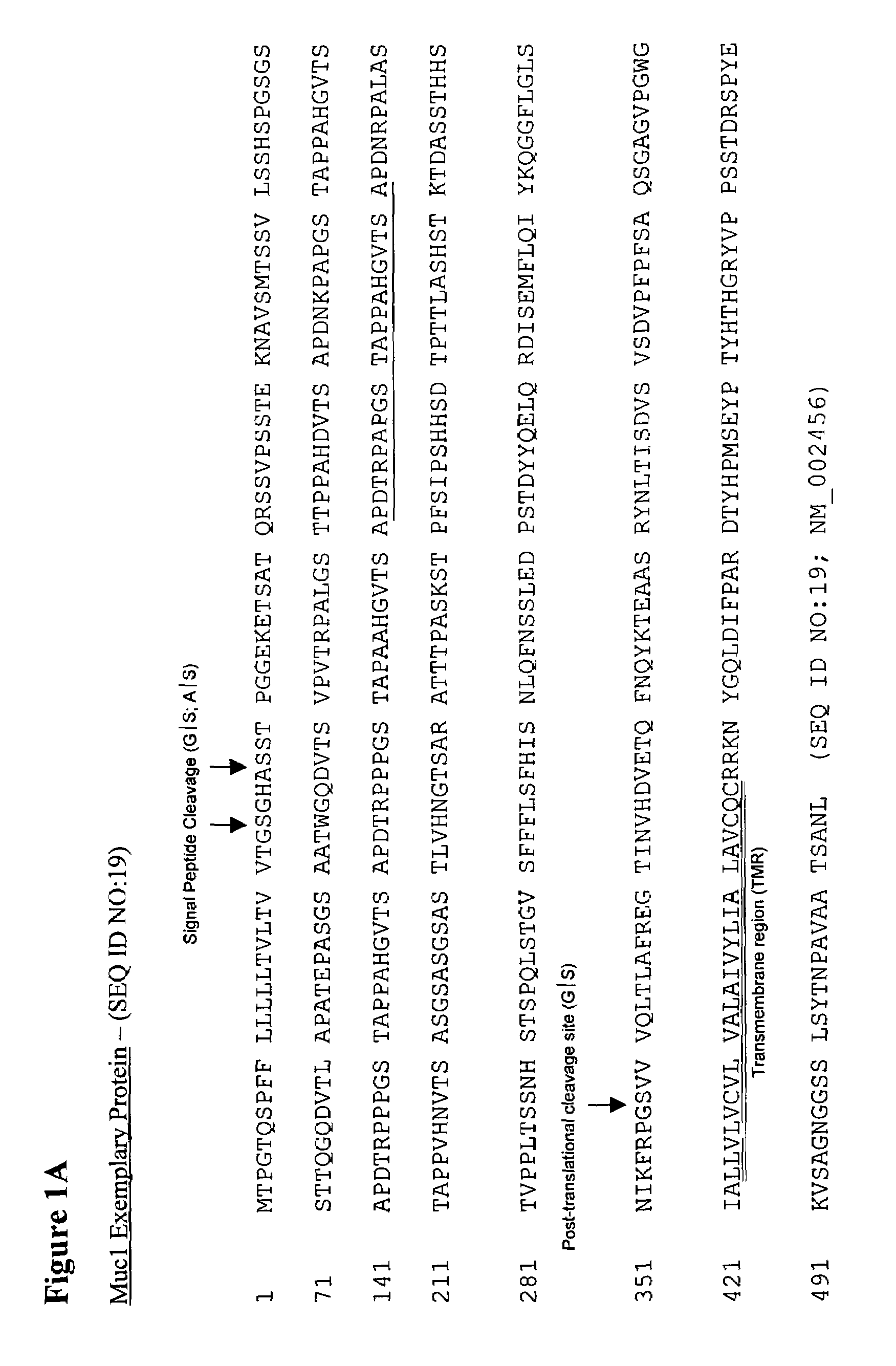
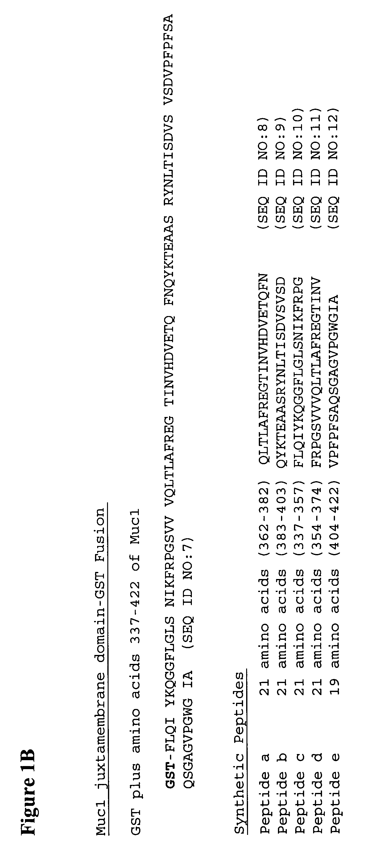
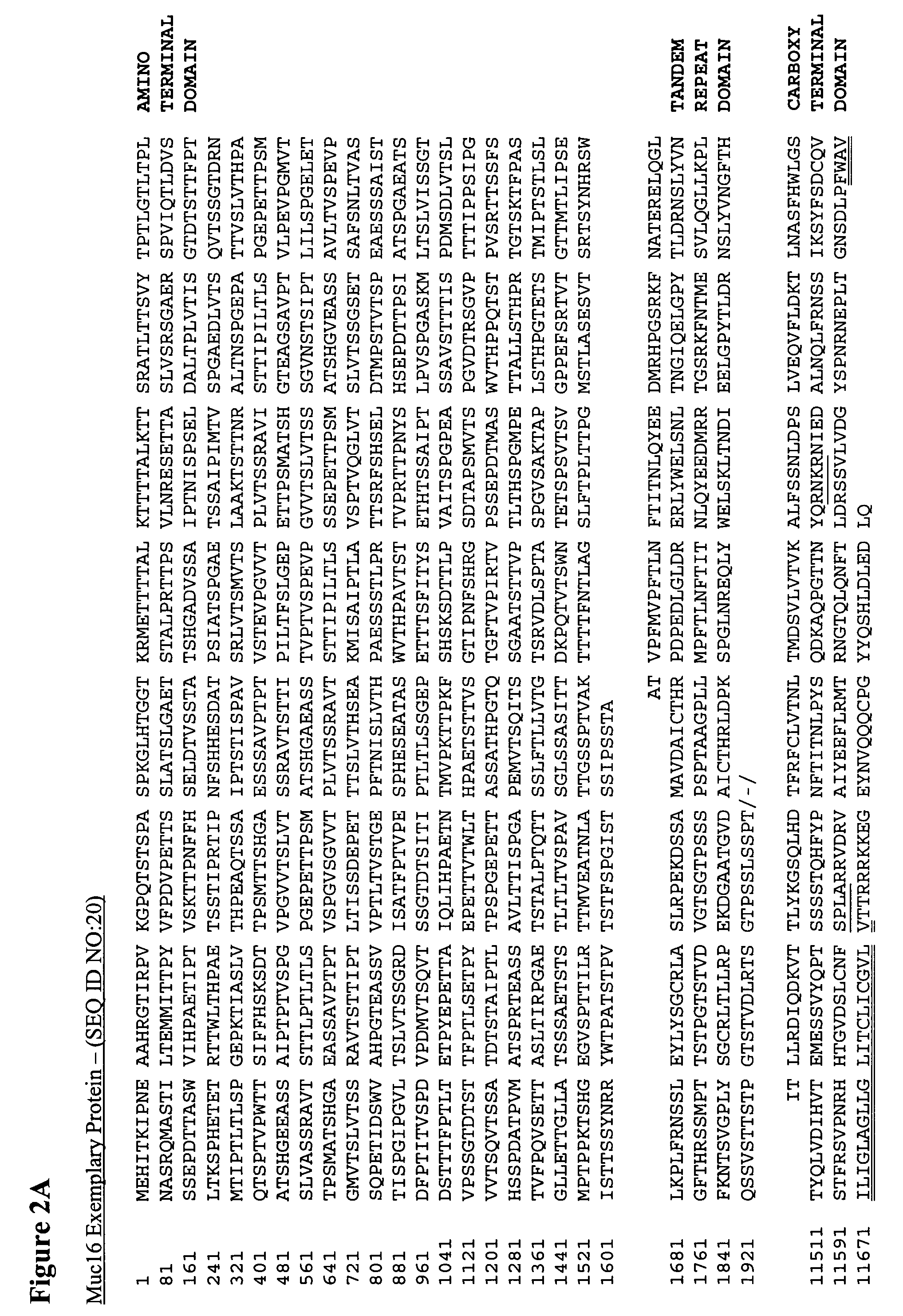
[0098]Panels of monoclonal antibodies (Mabs) were raised against putative non-shed extracellular epitope(s) of human Muc1 or Muc16 by immunizing mice with synthetic peptides (Boston BioMolecules, Inc.) representing 20 or 21 amino acid sequences selected from the extracellular juxtamembrane regions of these molecules. Specifically, Muc16 Peptide a, SSVLVDGYSPNRNEPLTGNS (SEQ ID NO: 14), representing residues 11644–11663 of CA125 (Muc16; SEQ ID NO:20); and Muc1 Peptide a, QLTLAFREGTINVHDVETQFN (SEQ ID NO:8), representing residues 362–382 of Muc1 (SEQ ID NO:19), were used to generate Mabs.
[0099]To enhance immune responses in mice, the synthetic peptides were conjugated with the carrier protein keyhole limpet hemacyanin (KLH) via a cysteine residue added to the amino termini of the peptides at the time of synthesis (Boston BioMolecules, Inc.) and mixed with complete or incomplete Freund's ad...
example 2
Screening of monoclonal antibodies to the extracellular cell-associated domains of Muc1 and Muc16 by ELISA
[0101]Peptide-specific antibodies in hybridoma supernatants were screened initially using a solid phase peptide ELISA in which a biotinylated preparation of the non-KLH-conjugated specific peptide (Boston BioMolecules, Inc.) was used as the capture antigen. Immulon H2B 96-well plates were coated with 250 ng per well (50 μl at 5 μg / ml) of NeutrAvidin (Pierce), in 0.5 M carbonate buffer, pH 10 for 4–6.5 hours at room temperature with rocking. The wells were washed twice with 300 μl per well of wash buffer (Tris Buffered Saline (TBS) / 0.1% Tween-20) and blocked with 200 μl per well of TBS / 3% BSA for 1 hour at room temperature with rocking. The biotinylated Muc1 Peptide a or Muc16 Peptide a were captured by the NeutrAvidin by incubation with 50 ng per well (50 μl of 1 μg / ml) of biotinylated peptide for 1 hour at room temperature (Muc16) or overnight at 4° C. (Muc1) with rocking. The ...
example 3
Screening of Monoclonal Antibodies to the Extracellular Cell-Associated Domains of Muc1 by Flow Cytometry
[0105]In addition to ELISA screening, the hybridoma supernatants were screened for binding to antigen positive tumor cell lines by flow cytometry. Muc1 hybridoma supernatants were screened using CaOV-3 cells and Muc16 hybridoma supernatants were screened using OVCAR-3 cells. For Muc1 screening, CaOV-3 cells were grown to 95% confluency on 15 cm tissue culture plates in complete media RPMI (Cambrex) supplemented with 10% heat-inactivated fetal bovine serum (Atlas), 50 Units / ml of penicillin / 50 μg / ml streptomycin (Cambrex), 2 mM L-Glutamine (Cambrex)) at 37° C. in 5% CO2. Cells were given 30 ml of fresh media one day before harvest. The cells were washed twice with phosphate buffered saline (PBS) and dissociated from the plate by incubation with 3 ml of Cellstripper (Mediatech, Inc.) at 37° C. for 10 minutes. The cells were washed in 20 ml of ice cold FACS Buffer (2% Goat Serum in ...
PUM
| Property | Measurement | Unit |
|---|---|---|
| Fraction | aaaaa | aaaaa |
| Volume | aaaaa | aaaaa |
| Volume | aaaaa | aaaaa |
Abstract
Description
Claims
Application Information