
Systems and methods for image processing of 2D medical images

a technology of image processing and medical images, applied in the field of image processing, can solve the problems of user division of attention, difficult to see where the image is located in the object, and difficult to interpret image slices

Active Publication Date: 2011-08-30
GENERAL ELECTRIC CO
View PDF14 Cites 41 Cited by
  • Summary
  • Abstract
  • Description
  • Claims
  • Application Information

AI Technical Summary

Benefits of technology

This approach enhances the understanding and interpretation of image slices by reducing the risk of orientation errors and computational complexity, providing a more intuitive and efficient method for image processing that maintains diagnostic quality without the need for extensive computational resources.

Problems solved by technology

Image slices are not easy to interpret.
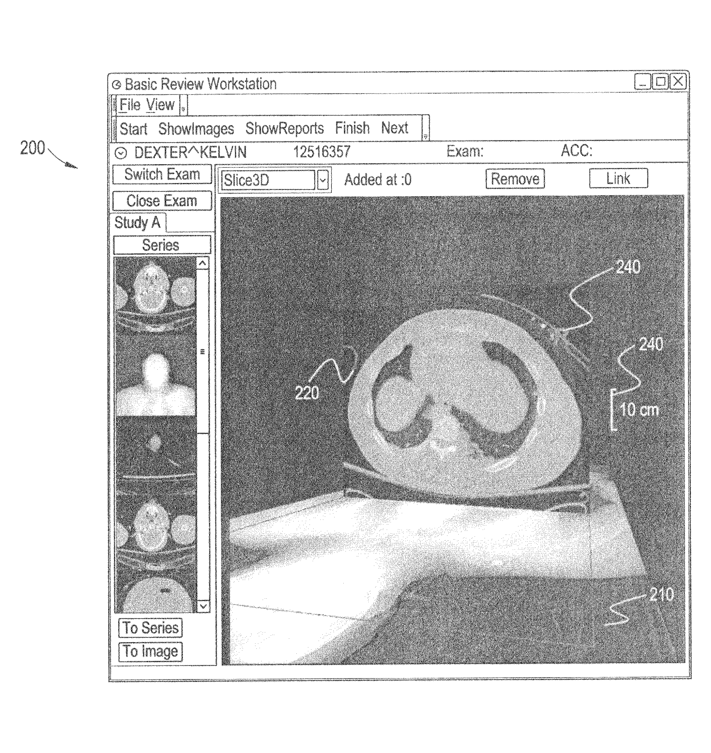
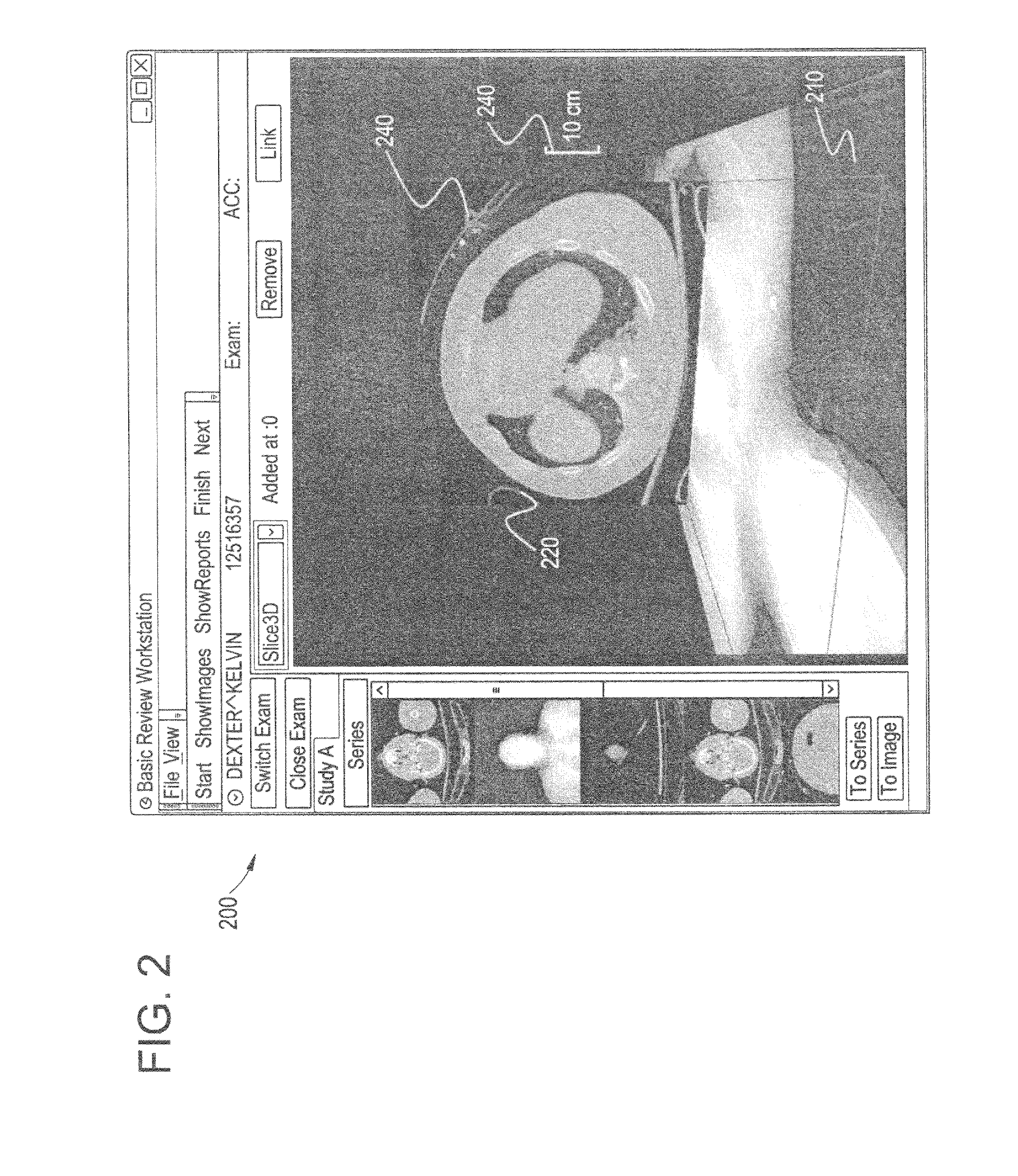
From a 2D image, it is difficult to see where the image is located in the object.
Although cut lines are useful, this method forces the user to divide their attention between two views.
Further, there is a risk of mistakes.
As another example, cut lines are not easy to understand by casual users, such as referring physicians and patients.
However, 3D image rendering requires 3D reconstruction which involves increased computational power and is slow to calculate.
This method also may be confusing to a user due to the amount of information that is displayed.
That is, a user may experience information overload.

Method used


Image

  • Systems and methods for image processing of 2D medical images
  • Systems and methods for image processing of 2D medical images
  • Systems and methods for image processing of 2D medical images

Examples


Embodiment Construction

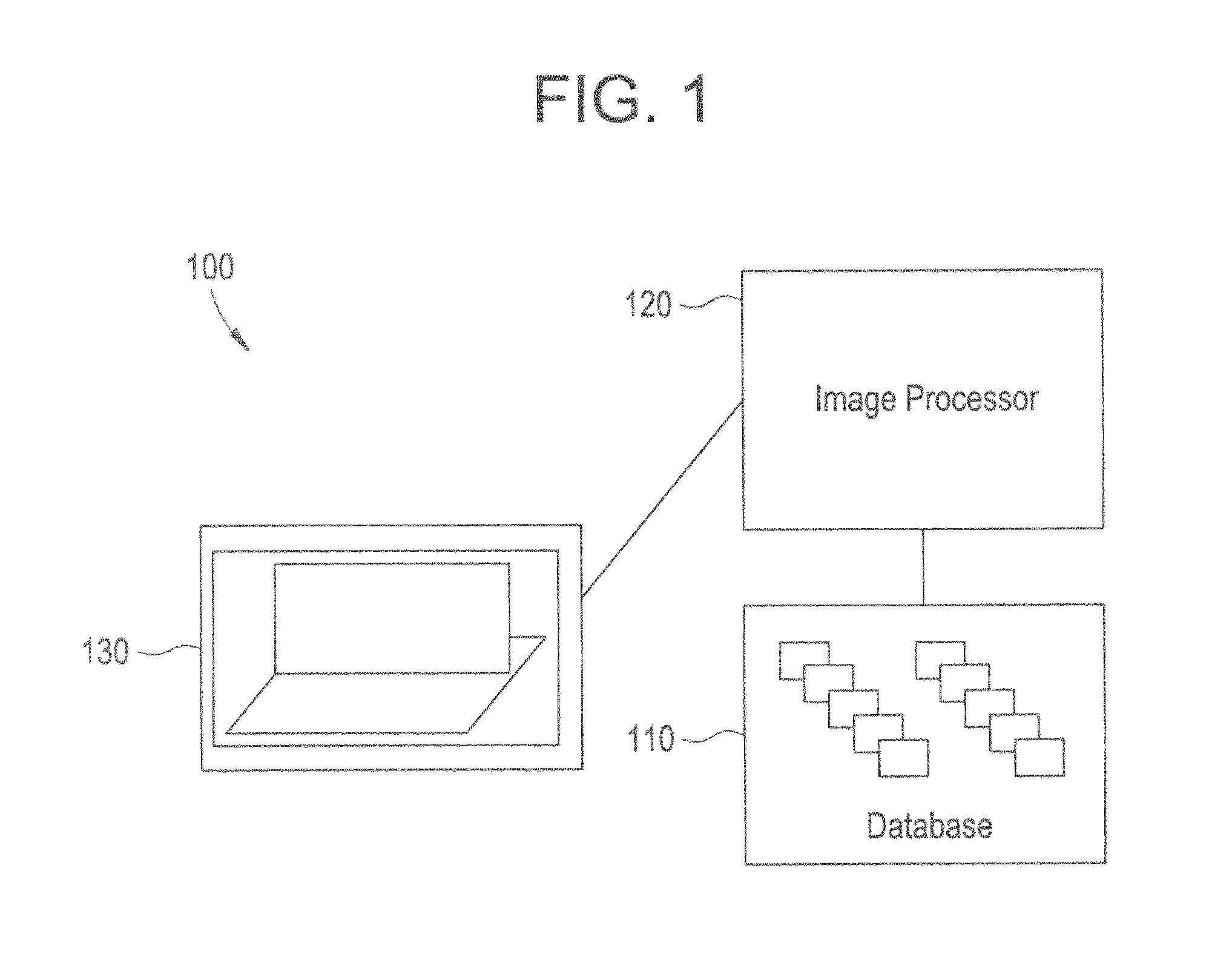
[0016]FIG. 1 illustrates a system 100 for image processing according to an embodiment of the present invention. The system 100 includes a database 110, an image processor 120, and a display 130.

[0017]The image processor 120 is in communication with the database 110 and the display 130.

[0018]In operation, the image processor 120 selects a selected image slice from the database 110. The image processor 120 generates a display image including a localizer image and the selected image slice. The display 130 displays the display image generated by the image processor 120.

[0019]The database 110 includes one or more image slices. These image slices may be taken using a scan of an object such as a patient, for example. For example, images slices may be acquired from an imaging system such as a magnetic resonance imaging (MRI), computed tomography (CT), or a Positron Emission Tomography (PET) modality.

[0020]The image processor 120 is adapted to select an image slice. The image slice may be se...


PUM


Abstract

Certain embodiments of the present invention provide a system for image processing including a database, an image processor, and a display. The database includes a plurality of image slices. Each image slice in the plurality of image slices is based at least in part on an object. The image processor is adapted to generate a display image. The display image includes a selected image slice rendered about perpendicular to a localizer image. The selected image slice is selected from the plurality of image slices. The display is adapted to display the display image.

Description

BACKGROUND OF THE INVENTION[0001]The present invention generally relates to image processing. More specifically, the present invention relates to systems and methods for image processing of two-dimensional (2D) medical images.[0002]Hospitals and other medical facilities, such as clinics and imaging centers, continually seek to improve or optimize utilization of resources and productivity. Such optimization may be achieved though faster or more understandable image processing. For example, improved image processing may include faster calculations, lower usage of computer hardware graphics, and lower storage requirements. Further, simplified images, better optimization of a single screen, and undistorted images may each improve productivity and can lead to decreased mistakes.[0003]Many techniques are used currently to process images involving image slices. Image slices are 2D images that may be created using a tomographic scan of an object, for example. In a tomographic scan, image sl...

Claims


Application Information

Patent Timeline
Patent Type & Authority Patents(United States)
IPC IPC(8): G06K9/00G09G5/00A61B8/00G16H30/40
CPCG06T7/0012G06T19/00G06T2200/24G06T2207/10072G06T2207/20108G06F19/321G16H30/40
Inventor DE VAAN, JAN ANNE NIELS
Owner GENERAL ELECTRIC CO
Learn More