Method for determining cardiac impulse conduction and associated medical device

a technology of cardiac impulse and cardiac impulse, applied in the field of cardiac impulse conduction determination, can solve the problems of difficult reconstruction of such a vector cardiogram, difficult diagnosis, and limited assessment of cardiac impulse conduction, and achieve the effects of improving the identification of individual electrodes, and reducing the difficulty of diagnostic errors

Inactive Publication Date: 2011-10-11
SIEMENS HEALTHCARE GMBH
View PDF7 Cites 3 Cited by
  • Summary
  • Abstract
  • Description
  • Claims
  • Application Information

AI Technical Summary

Benefits of technology

[0024]Through direct determination of the positions of the ECG electrodes from the image recording or the image recordings, electrode capture and / or detection is possible without additional conversion procedures. It is thus no longer necessary to carry out a complicated conversion or transformation which is based on image recordings produced previously or independently of the ECG recording and which possibly do not show the electrodes at all or not in the currently correct position for the ECG recording.
[0050]The medical device is thus configured such that, through a combination of ECG data and data of an imaging device, for example a magnetic resonance device, the information about the electric potentials at the body surface of a patient can be acquired simultaneously with image data which provides the shape or anatomy of the heart and of the thorax and the position of the electrodes. The acquisition of all the information that is necessary for a comprehensive reconstruction of the propagation of excitation in the heart of the patient, in particular as a 3D-vector cardiogram, is thus made possible, in particular through a simultaneous or ECG-triggered recording of potential data and image data.

Problems solved by technology

In a conventional ECG, assessment of cardiac impulse conduction is restricted, however, as the electrodes even in a 12-channel ECG are not arranged in all planes, so that propagations of the electric field which have a vector perpendicular to the planes captured are not captured.
As a result of this, however, diagnosis becomes increasingly difficult as the number of curves is very high and these are ultimately difficult to interpret so that highly trained specialists are required to assess them.
In a living patient, the reconstruction of such a vector cardiogram is possible only with very great difficulty as the currently existing solutions are associated with considerable costs.
The reason why the currently existing methods for the reconstruction of vector cardiograms are very costly and of limited practicability, is that, in order to reconstruct the propagation of excitation of the heart on the basis of the measured potentials, the precise position of the electrodes used and the anatomy, i.e. in particular the shape of the heart, have to be known.

Method used


Image

  • Method for determining cardiac impulse conduction and associated medical device
  • Method for determining cardiac impulse conduction and associated medical device
  • Method for determining cardiac impulse conduction and associated medical device

Examples


Embodiment Construction

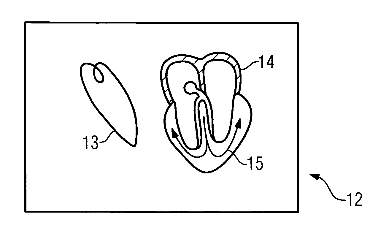
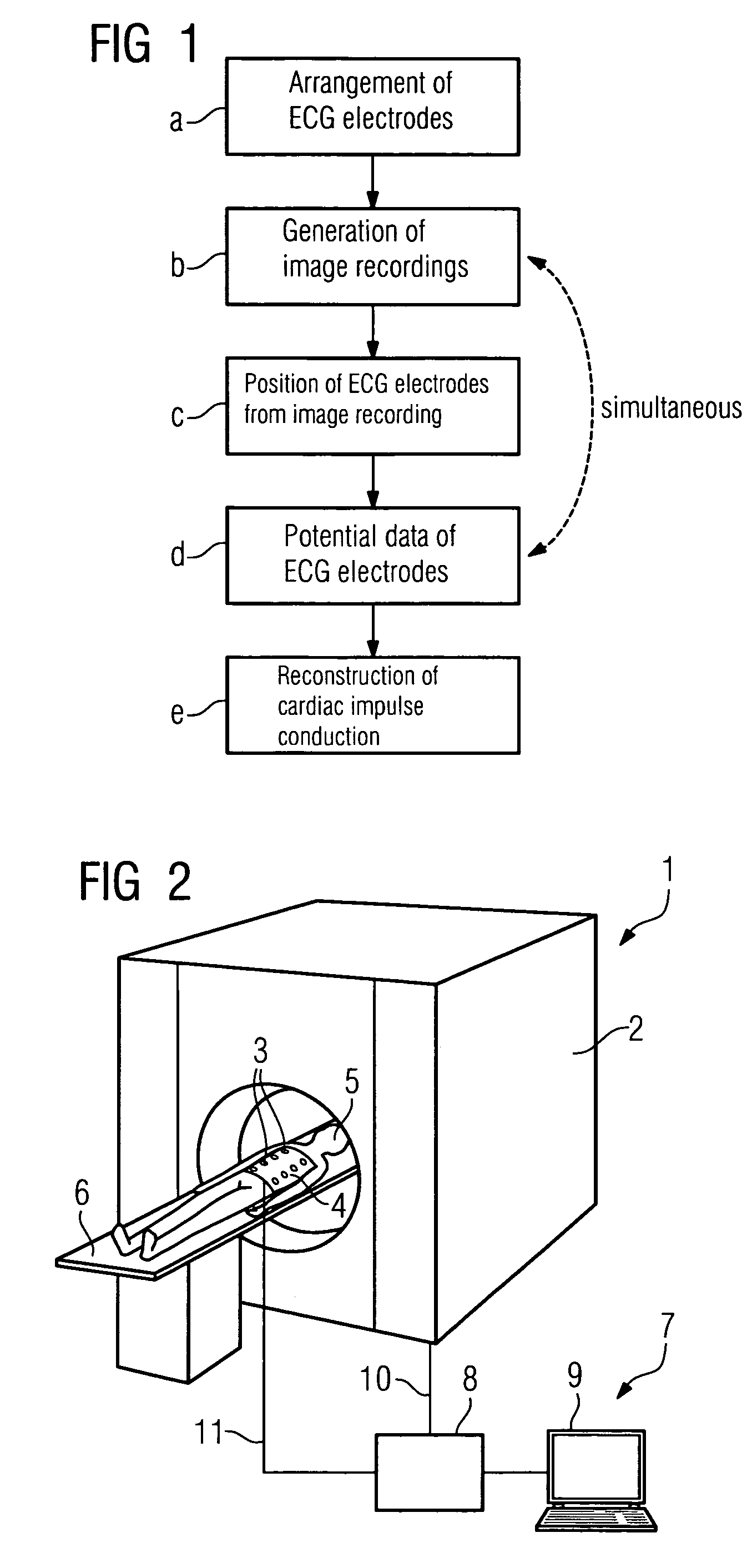
[0066]FIG. 1 represents a flow diagram of a method according to the invention comprising steps a-e. Firstly, in step a, the ECG electrodes for recording the electric potentials are arranged on the upper body of the patient. Then, in accordance with step b, at least one image recording or even a film of image recordings of at least one area of the body of the patient, advantageously of the area in which the ECG electrodes are arranged, is generated. This may, for example, be carried out by means of a magnetic resonance tomograph or by means of another imaging device which records e.g. the thorax of the patient.

[0067]The positions of the ECG electrodes are, in accordance with step c, determined directly from the image recording. Consequently, not only is the anatomy, i.e. the shape of the heart and of the thorax of the patient, derived from the image recording, but the position of the electrodes is also determined therefrom.

[0068]In step d, a recording of potential data of the ECG ele...


PUM


Abstract

A method for determining cardiac impulse conduction, in particular three-dimensional cardiac impulse conduction, in a patient, comprising: generating an image recording of an area of the body of the patient capturing at least partially electrocardiogram electrodes arranged on the body of the patient by an imaging modality; determining positions of the electrocardiogram electrodes in a system of coordinates assigned to the imaging modality; recording of potential data of some of the electrocardiogram electrodes; and reconstructing cardiac impulse conduction depending on the determined positions of the electrocardiogram electrodes, the image recording and the recording of potential data of the electrocardiogram electrodes, wherein at least one image recording is generated substantially simultaneously with the recording of potential data of the electrocardiogram electrodes or is generated in the period between two recordings of potential data of the electrocardiogram electrodes.

Description

CROSS REFERENCE TO RELATED APPLICATIONS[0001]This application claims priority of German application No. 10 2007 007 563.6 filed Feb. 15, 2007, which is incorporated by reference herein in its entirety.FIELD OF THE INVENTION[0002]The invention relates to a method for determining cardiac impulse conduction, in particular for determining three-dimensional cardiac impulse conduction, in a patient and to an associated medical device for determining cardiac impulse conduction.BACKGROUND OF THE INVENTION[0003]In numerous disorders in the area of cardiology, for example in a case of myocardial infarction or in cases of cardiac arrhythmia, the propagation of electrical signals in the heart is altered. This results in a change in the electric field of the heart, the sum vector of which is evident in changes in the electric potentials on the body surface of a patient.[0004]In an electrocardiogram (ECG), the electric potentials and / or the changes in the electric potentials at the body surface o...

Claims


Application Information

Patent Timeline
Patent Type & Authority Patents(United States)
IPC IPC(8): A61B5/0402
CPCA61B5/0402A61B6/5247A61B8/4416A61B5/7285A61B6/503A61B6/541Y10S128/922A61B2019/5454Y10S128/92A61B2090/3954A61B5/318A61B5/367
Inventor SCHMIDT, SEBASTIAN
Owner SIEMENS HEALTHCARE GMBH
Why Eureka
Try Eureka