Biological surgical patch and method of making

a biochemical and surgical technology, applied in the field of surgical patches, can solve the problems of difficult control of the degradation of these materials, inability to cure non-bacterial inflammatory diseases, and inability to achieve the effect of good biocompatibility and safe/reliable application

Active Publication Date: 2012-01-24
SUMMIT GD BIOTECH
View PDF57 Cites 0 Cited by
  • Summary
  • Abstract
  • Description
  • Claims
  • Application Information

AI Technical Summary

Benefits of technology

[0008]It is an object of the present invention to provide a biological surgical patch having no immune rejection with good biocompatibility and safe / reliable application, and a method of preparation thereof.
[0009]In order to accomplish the objects of the present invention, the present invention provides a biological surgical patch made by a method that includes the steps of providing a natural animal tissue that has a substrate, crosslinking and fixing the substrate, minimizing the antigens from the substrate, tanning the substrate, and incorporating an active layer in the substrate.

Problems solved by technology

These materials often lead to non-bacterial inflammatory diseases due to physical irritation and complications caused by chronic rejective reaction.
However, the rate of degradation of these materials is difficult to control to coincide with the rate of tissue recovery, so the efficacy is often uncertain due to rapid degradation.
In addition, the degraded products can bring local acidity which affects normal healing of the repaired tissues.
Animal membrane tissues such as fascia and bovine pericardium have also been utilized in recent years through conventional processes including defatting, cell removal and fixation with glutaraldehyde, but elimination of antigens is hardly effective because cell removal is employed as the only means for eliminating antigens.
Additionally, fixation of tissues with glutaraldehyde is achieved by crosslinking through acetalization, and glutaraldehyde is released during degradation, leaving residual toxicity and causing more potent cellular toxicity so that its growth in the inherent tissue becomes difficult, resulting in poor repair effect.

Method used


Image

  • Biological surgical patch and method of making
  • Biological surgical patch and method of making
  • Biological surgical patch and method of making

Examples


example 1

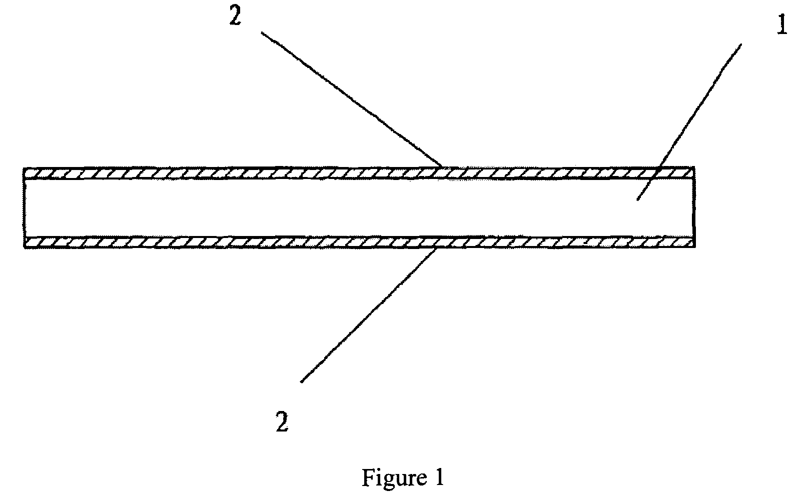
[0039]As shown in FIG. 1, the biological surgical patch comprises (i) a substrate 1 prepared from porcine or bovine pericardium by crosslinking fixation with a non-aldehyde fixative, eliminating antigens and improving the strength with a protein, and (ii) active surface layers 2 formed by on both the top and bottom surfaces of substrate 1 by coupling an active component such as a specific polypeptide or glycosaminoglycan. One example of the polypeptides is the polypeptide obtained from the condensation of 16 lysines (K16), glycine (G), arginine (R), asparagic acid (D), serine (S), proline (P) and cysteine (C), and the glycosaminoglycan is hyaluronic acid, chondroitin sulfate, dermatan sulfate, heparin, acetylheparin sulfate or keratan sulfate.

[0040]The method of preparation of the biological surgical patch of the present invention includes the following steps, using porcine or bovine pericardium as the substrate:

[0041]1. Selection of materials and pretreatment: Fresh porcine or bovi...


PUM


Abstract

A biological surgical patch made by a method that includes the steps of providing a natural animal tissue that has a substrate, crosslinking and fixing the substrate, minimizing the antigens from the substrate, tanning the substrate, and incorporating an active layer in the substrate.

Description

BACKGROUND OF THE INVENTION[0001]1. Field of the Invention[0002]The present invention relates to a medical prosthesis for human implantation, and in particular, to a surgical patch that is used for repairing tissues or organs during surgery.[0003]2. Description of the Prior Art[0004]Repair of defective tissues and organs is often required in modern surgical techniques, for example, repair of dura mater, repair of defective pleura, repair of peritoneum, repair of hernial strain, repair of diaphragm, repair of blood vessels, repair of atrial septum, repair of pericardium, repair of kidneys, etc. There are many “surgical sticking patches” that are currently being provided to meet clinical demands. However, these products were first prepared with synthetic materials.[0005]These synthetic materials utilized in preparing the surgical sticking patches include polypropylene, polyethylene, polyamide, Dacron resin, polytetrafluoroethylene, silicone gel, and carbon fiber, among others, which a...

Claims


Application Information

Patent Timeline
Patent Type & Authority Patents(United States)
IPC IPC(8): A61F2/02A61K35/34
CPCA61L27/3604A61L27/3683
Inventor XU, GUO-FENG
Owner SUMMIT GD BIOTECH
Why Eureka
Try Eureka