Automatic alignment of magnetic resonance imaging (MRI) brain scan by anatomic landmarks

a magnetic resonance imaging and brain scan technology, applied in the field of automatic brain scanning, can solve the problems of incongruous anatomy on diagnostic images, patient must remain absolutely, manual process may take several seconds, etc., and achieve the effect of minimizing differences and maximizing image gradient magnitud

Inactive Publication Date: 2012-05-29
SIEMENS HEALTHCARE GMBH
View PDF4 Cites 25 Cited by
  • Summary
  • Abstract
  • Description
  • Claims
  • Application Information

AI Technical Summary

Benefits of technology

[0009]The step of identifying an MSP line in each of the coronal and transverse views comprises: detecting a position of the patient's head by fitting an ellipse to each of the coronal and transverse views that maximizes an image gradient magnitude in a boundary region of the ellipse; establishing a symmetrical axis which minimizes a difference between the coronal and transverse views on either side of the symmetrical axis; characterizing low intensity pixels in the coronal and transverse views by using smoothed local differential operators; finding pixels in the coronal and transverse views that form a space between hemispheres in each of the coronal and transverse views; and running a linear regression with robust weights to define a line in each of the coronal and transverse views that best separates the two hemispheres, wherein the line is the MSP line.
[0021]The step of identifying an MSP line in each of the coronal and transverse views comprises: detecting a position of the patient's head by fitting an ellipse to each of the coronal and transverse views that maximizes an image gradient magnitude in a boundary region of the ellipse; establishing a symmetrical axis which minimizes a difference between the coronal and transverse views on either side of the symmetrical axis; characterizing low intensity pixels in the coronal and transverse views by using smoothed local differential operators; finding pixels in the coronal and transverse views that form a space between hemispheres in each of the coronal and transverse views; and running a linear regression with robust weights to define a line in each of the coronal and transverse views that best separates the two hemispheres, wherein the line is the MSP line.

Problems solved by technology

Even when the same anatomic landmarks are used, due to inter- and intra-operator variation, the scan can be executed in different ways, resulting in inconsistent anatomy on diagnostic images.
In addition, the manual process may take several seconds, during which the patient must remain absolutely still.
This is inconvenient for the patient (many of whom find the machine claustrophobic) and ties up a very expensive piece of equipment.

Method used


Image

  • Automatic alignment of magnetic resonance imaging (MRI) brain scan by anatomic landmarks
  • Automatic alignment of magnetic resonance imaging (MRI) brain scan by anatomic landmarks
  • Automatic alignment of magnetic resonance imaging (MRI) brain scan by anatomic landmarks

Examples


Embodiment Construction

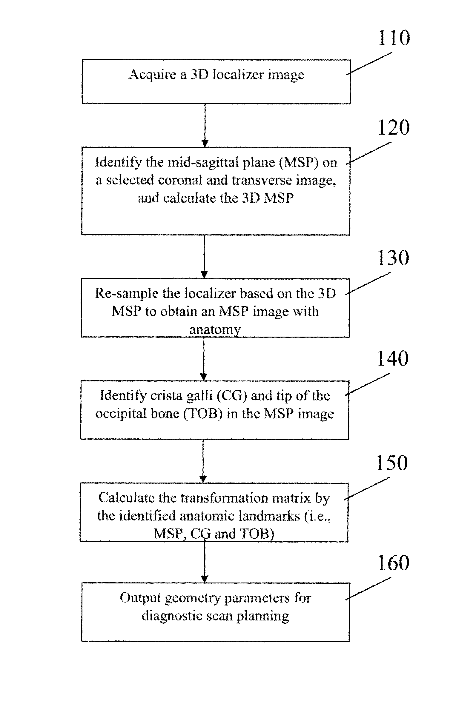
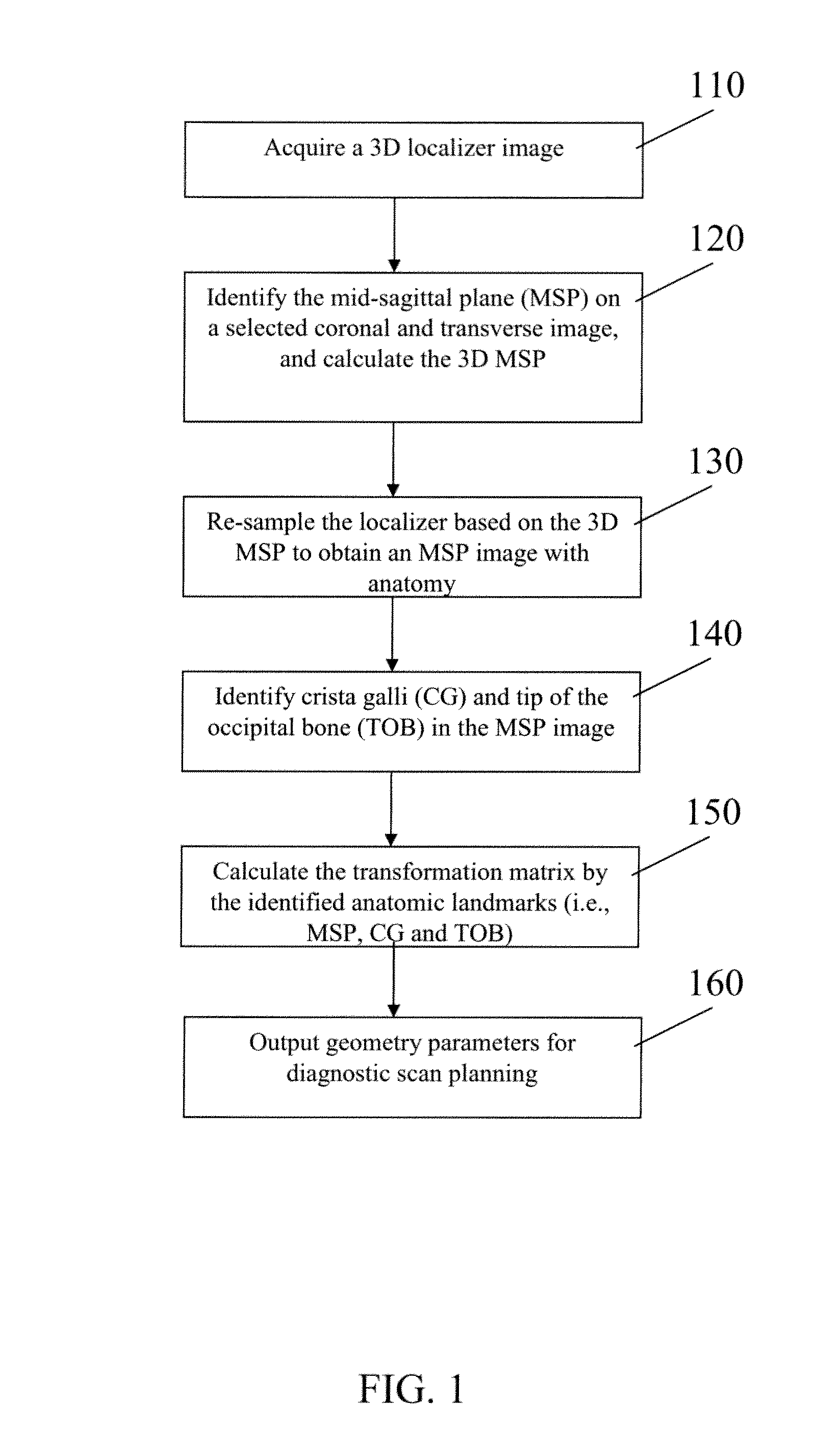
[0034]A method to automatically align brain scans for diagnostic scan planning, in accordance with an exemplary embodiment of the present invention, will now be described with reference to the accompanying drawings.

[0035]The method requires a three-dimensional (3D) localizer to start the scan planning. The fully automated process provides standardized and reproducible brain scanning for different hospitals, departments and operators, in order to perform disease diagnosis, treatment evaluation and therapy control.

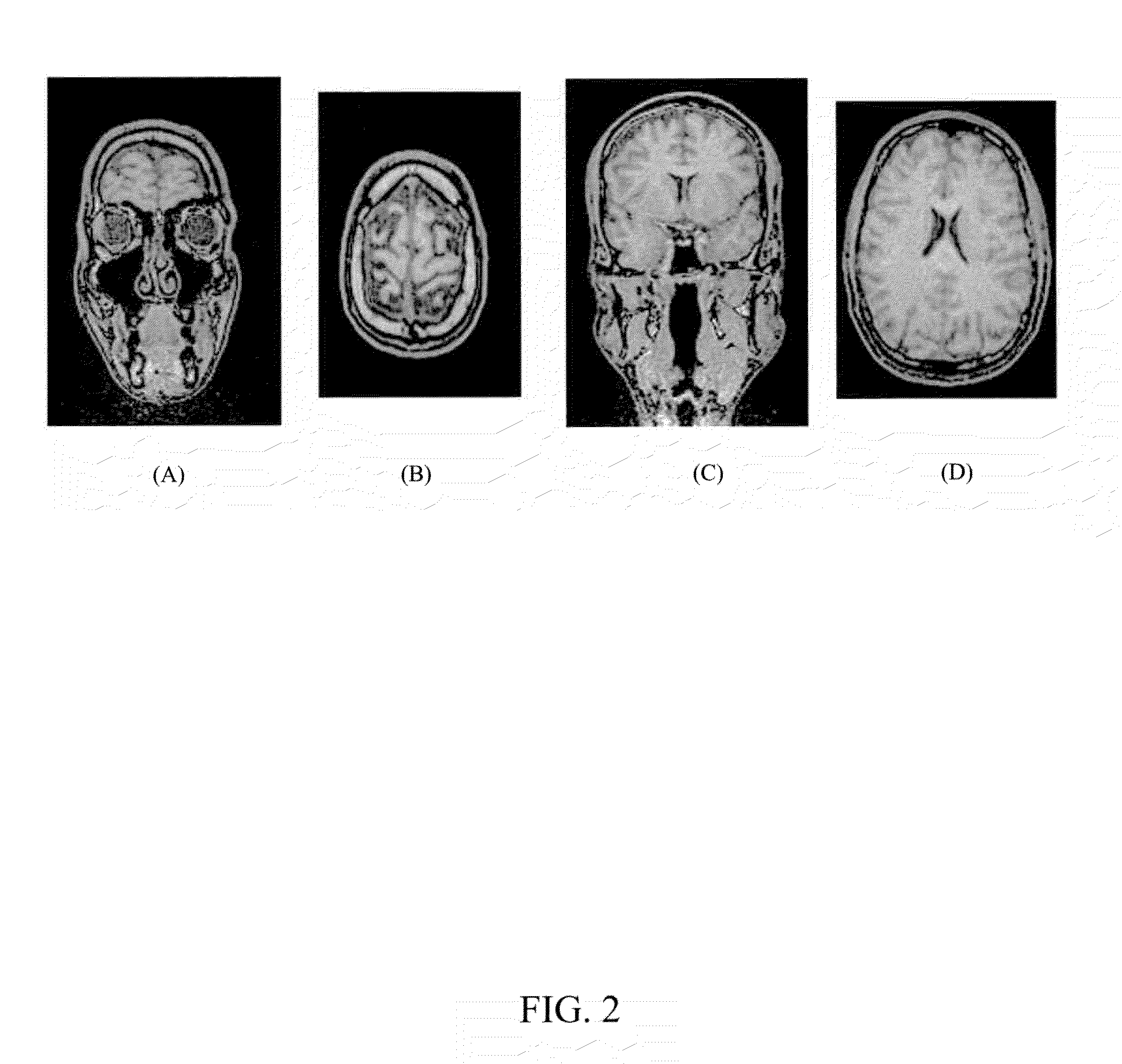
[0036]The automatic brain alignment algorithm starts with a dedicated three-dimensional (3D) localizer (110). From the 3D localizer, a coronal and a transverse image are selected to identify the mid-sagittal plane (MSP) and the 3D MSP is calculated from the MSP detection results on the selected coronal and transverse images (120). Then the original 3D localizer volume is re-sampled based on the orientation of the MSP to obtain an image with MSP anatomy (130). On the MSP imag...


PUM


Abstract

A method to automatically align magnetic resonance (MR) brain scans for diagnostic scan planning, including: acquiring a three-dimensional (3D) localizer image of a patient; selecting a two-dimensional (2D) coronal view and a 2D transverse view from the localizer image; identifying a mid-sagittal plane (MSP) line in each of the coronal and transverse views and calculating a 3D MSP based on the MSP lines; reconstructing the localizer image based on an equation for the 3D MSP to obtain an image of the MSP of the patient's brain; identifying crista galli (CG) and tip of the occipital bone (TOB) in the image of the MSP of the patient's brain; calculating a transformation matrix based on the MSP, CG and TOB in the image and using the transformation matrix to obtain a scan plan for the patient; and outputting the scan plan for the patient.

Description

CROSS-REFERENCE TO RELATED APPLICATION[0001]This application claims the benefit of U.S. Provisional Application No. 60 / 977,488, filed Oct. 4, 2007, the disclosure of which is incorporated by reference herein in its entirety.BACKGROUND OF THE INVENTION[0002]1. Technical Field[0003]The present invention relates to automating brain scanning.[0004]2. Discussion of the Related Art[0005]A typical brain scanning process in most hospitals starts with localizer or scout images acquired by fast MR sequences. This produces a low-resolution image indicating the patient's position within the machine. Then an operator plans a diagnostic scan sequence, which takes a longer time, from the localizer images using anatomic landmarks. However, for different hospitals, departments and operators, different anatomic landmarks may be used to plan the scanning. Even when the same anatomic landmarks are used, due to inter- and intra-operator variation, the scan can be executed in different ways, resulting in...

Claims


Application Information

Patent Timeline
Patent Type & Authority Patents(United States)
IPC IPC(8): A61B5/05
CPCA61B5/055A61B5/417A61B5/4504
Inventor ZHANG, LINOVAK, CAROL L.XU, QINGCHEN, CHONG
Owner SIEMENS HEALTHCARE GMBH
Why Eureka
Try Eureka