Laparoscopic trainer and method of training

a trainer and laparoscopic technology, applied in the field of surgical training, can solve the problems of high cost of laboratory animals for surgical training, surgeons are not able to rely on their sense of feel and direct tactile response during laparoscopic surgery, and achieve the effect of low cos

Inactive Publication Date: 2013-06-11
WANG SHYH JEN +1
View PDF6 Cites 53 Cited by
  • Summary
  • Abstract
  • Description
  • Claims
  • Application Information

AI Technical Summary

Benefits of technology

[0024]According to one aspect of the present invention, the surgery trainer further provides a stopper for each trocar or reducer cap to prevent it be pulled out. The stopper has an inside dimension to be fitted on the trocar or reducer cap and an outside dimension, which is greater than the size of aperture so that the trocar or reducer cap would not be pulled out the membrane after fitting with the stopper.
[0026]The present invention provides an endoscopically surgical trainer that realistically simulates a surgeon's view of an laparoscopic surgery site at a low cost. According to one embodiment of the present invention, the laparoscopic surgery trainer includes a camera handle passing through the aperture on the face wall. The camera, is installed in front of the handle to catch the image of a simulated anatomical structure on the base. Following, the image signal can be transmitted to and shown on a screen. The feature of image displayed is very similar to that of real laparoscopic system.
[0027]According to another embodiment of the invention, the surgery trainer further provides a plain spherical bearing set on the face wall. Thereby, the camera can be adjusted in and out as well as different orientation angles. Furthermore, the handle can locked in place so that the trainees can perform the trainer by herself / himself without the help by others.
[0028]According to another aspect of the present invention, the routine skills such as laparoscopic suturing and knot tying can be repeatedly practiced by trainees on a low-cost laparoscopic surgery trainer. The present invention thus provides an opportunity for training to develop realistic laparoscopic surgical skills.

Problems solved by technology

However, unlike the conventional techniques, the surgeon is not able to rely on his or her sense of feel and direct tactile response during laparoscopic surgery.
Further, visual response is limited to the two-dimensional image viewed through the laparoscopic video output, in which many structures may look similar.
The use of laboratory animals for surgical training is very expensive and is sometimes also subject to popular debate.
Additionally, animal specimens have a short viability on the operating table, and thus provide the trainee with a limited period of time in which to practice repeated techniques.
Though these devices have prominent features, they also have significant drawbacks.
Although this simulator functions satisfactorily in most cases, it tends to suffer from several problems which limit its usefulness.
One such drawback is that, if encourages the use of Pelviscope.
Endoscopes are expensive and are not readily available to many surgeons much less to the initiates.
Yet another drawback of the Pelvi-Trainer is that, when the surgeon uses the Pelviscope, she / he actually carries out the simulated instrumentation and practice procedures under direct monocular vision.
Still another drawback of this simulator is that, it has only one plastic panel with multiple openings that are positioned primarily for practice procedure on the pelvic cavity.
Additional drawback of the Pelvi-Trainer is that, in the practice of simulation, the trainee can only use human tissue like placenta or animal parts.
These materials are not readily available and their procurement might be difficult.
One drawback that is very evident is that, the trainee has to use the apparatus with the aid of a scope which provides a magnified direct monocular vision similar to the Semm Pelvi-Trainer.
Yet another drawback of this apparatus is that, it has provision only for practice simulation on materials like animal parts or even human tissue which are suspended from the top plexiglass panel by chain beads and clips, a method exactly similar to the Semm Pelvi-Trainer.
The top lid plexiglass of the McKeown apparatus has multiple puncture positions that are appropriately used for simulation practice exercise on limited organ location, i.e., the placement and location of the apertures on the top lid is not suitable for practice simulation of laparoscopic surgery on the chest or in the region of the kidneys and this is also a drawback of the device.
Still another drawback; of this training apparatus is that, the trainee must always use an endoscope to perform practice simulation.
The endoscope is an expensive item and not readily available to most trainees, therefore the use of such apparatus is limited to hospital setting where the endoscope might be accessible to the trainee.
Another drawback of this device is that, the mounting of the organ object being practiced on is by clamps on bead chains suspended from the top plexiglass panel which makes the object a moving target and would need a steady hand to perform simulated delicate sewing technique.
It may result in learning a difficult but not necessary technique.
Furthermore, the smaller circular pressure sensitive plate, reinforcing the holding strength of plate against the in and out movement of the trocars, may still cause the trocars to be pulled out.
However, the inventor of the U.S. Pat. No. 5,722,836 only mentioned the concept, but not enabled the desire so to leave the trocar stop, flexible elastomeric cover and trocar be not shown in his specification.
However, even with the membrane with flexibility, the training realism is still fallen behind in that the membrane material always would not provide radial movement.
However, the problems of support wall without flexibility and trocars to be fallen out still existed in the U.S. Pat. No. 6,887,082.

Method used


Image

  • Laparoscopic trainer and method of training
  • Laparoscopic trainer and method of training
  • Laparoscopic trainer and method of training

Examples


Embodiment Construction

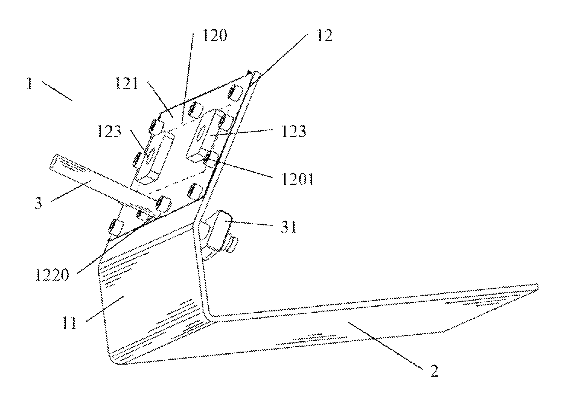
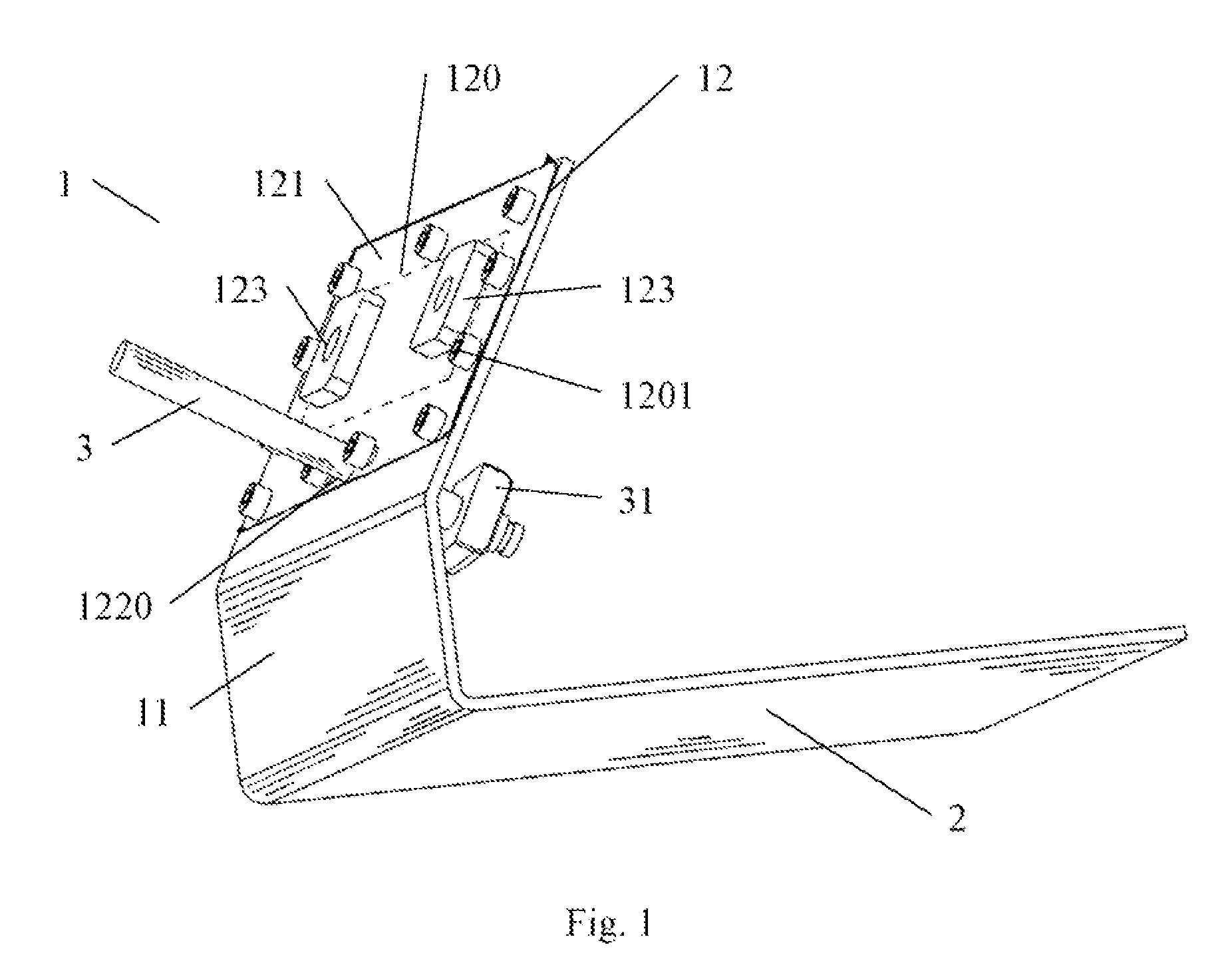
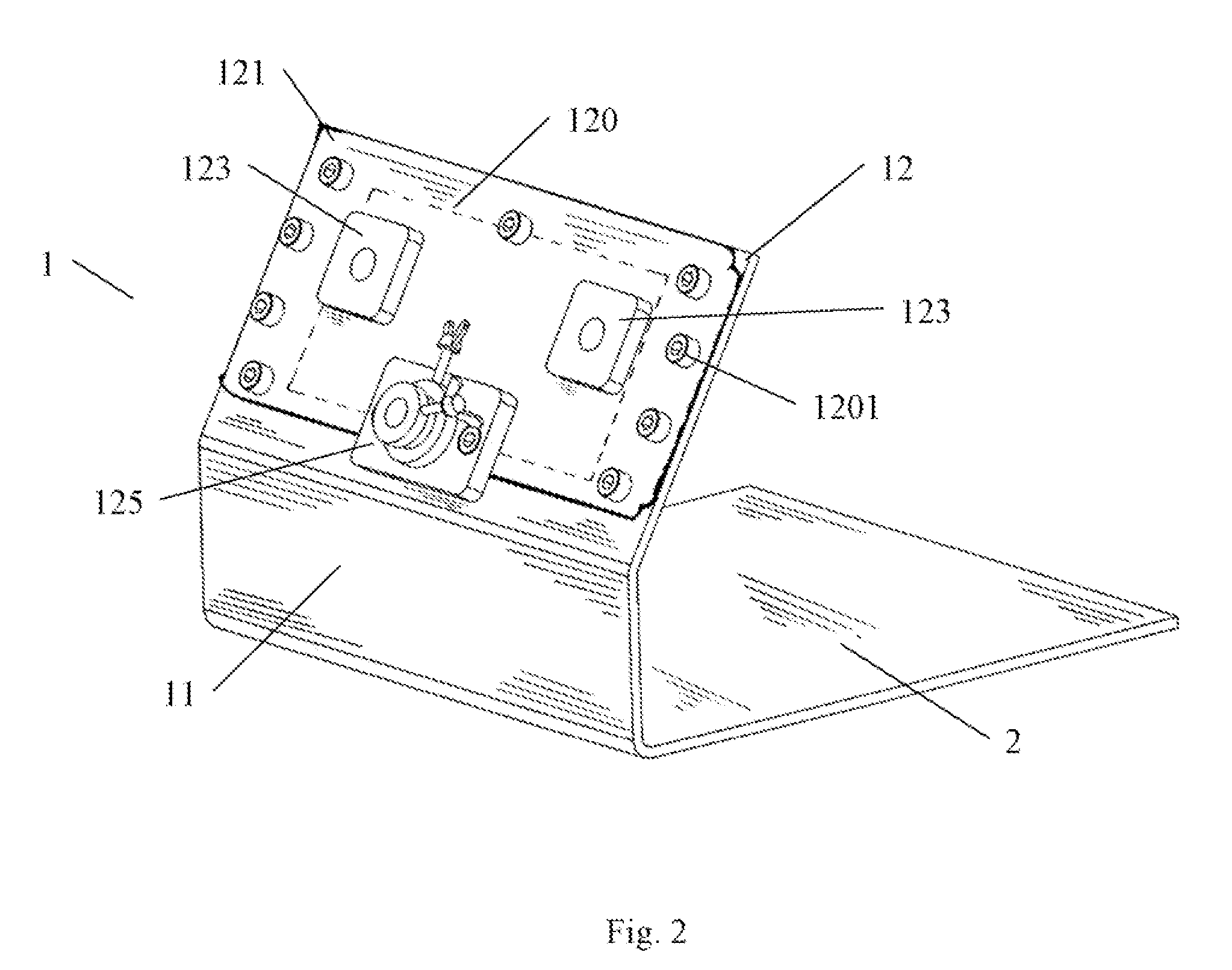
[0036]A trainer for surgeons to learn and practice laparoscopic surgical procedures is depicted generally in FIG. 1. The device comprises a face wall 1 and base 2. The base 2 is arranged to receive an operable structure. The face wall 1 is extended upwardly from a side of the base 2. The face wall 1 and base 2 both are basically formed from an opaque material. It will be appreciated that in other embodiments, the base 2 and a front wall 11, extended upwardly from the base 2, of the face wall 1 could be also formed from a transparent material. Extending upwardly and inwardly from the upper edge of the front wall 11 is an upper wall 12 of the face wall 1. The angle of the upper wall 12 relative to front wall 11 is greater than 90 degree. The angle is frequently set between 110 degree and 165 degree. The preferable angle is around from 120 degree to 160 degree. The most preferable angle is around from 130 degree to 150 degree. The position and orientation of the upper wall 12 of the fa...


PUM


Abstract

A surgical training device for the practice of surgical techniques comprises a base and a face wall extending from a side of said base such to block direct visualization of the operation area from the normal external position of the eyes of the trainee using the device. The face wall comprises a front wall and an upper wall. The front wall is extended upwardly from the base; the upper wall is then extended upwardly and inwardly from the front wall. A flexible membrane is stretched on upper wall and has at least two portals such that adaptors can be inserted on flexible membrane. The adaptors are further fitted with stoppers to prevent adaptors been pulled out from the membrane during training. The surgical instruments can pass through the openings of adaptors to manipulate simulated tissue or another operable structure on the base. A plain spherical bearing set, including a bearing seat and a plain spherical bearing, is also installed on said upper wall. The bearing seat has a bearing hole to interference fit with plain spherical hearing and two bolt holes to hold bearing seat on upper wall such that a camera handle can pass through plain spherical bearing to catch the image on base.

Description

FIELD OF THE INVENTION[0001]The present invention relates to surgical training, and particularly to a structure for use in learning laparoscopic surgical techniques.BACKGROUND OF TOE INVENTION[0002]In recent years, many invasive surgical and operative medical procedures have been developed utilizing laparoscopic techniques. The laparoscopic can reduce the trauma and recuperative periods associated with prior conventional surgical techniques. To date, the laparoscopic procedures have been developed for a wide variety of gynecological, chest, abdomen and urologic procedures, to place feeding tubes into the digestive tract, to take biopsies, to inspect for tumors, and to correct some types of infertility, by way of example. Additional techniques may be developed including laparoscopic techniques for sinus surgery, esophagus repair, fetal surgery and prophylactic breast cancer surgery. Basically, the laparoscopic entails inserting instruments through a small incision into an operative c...

Claims


Application Information

Patent Timeline
Patent Type & Authority Patents(United States)
IPC IPC(8): G09B23/28
CPCG09B23/285
Inventor WANG, SHYH-JENCHIN, TAIWAI
Owner WANG SHYH JEN
Why Eureka
Try Eureka