Methods for collecting and processing autografts, processed autografts, kits for collecting and transporting autografts, and tools for preparing autografts
a technology for autografts and autograft kits, applied in the field of methods for collecting and processing autografts, processing autografts, kits for collecting and transporting autografts, and tools for preparing autografts, can solve the problems of difficult preparation of synthetic implants, unpredictable clinical and aesthetic outcomes, and time-consuming and expensive problems
- Summary
- Abstract
- Description
- Claims
- Application Information
AI Technical Summary
Problems solved by technology
Method used
Image
Examples
example 1
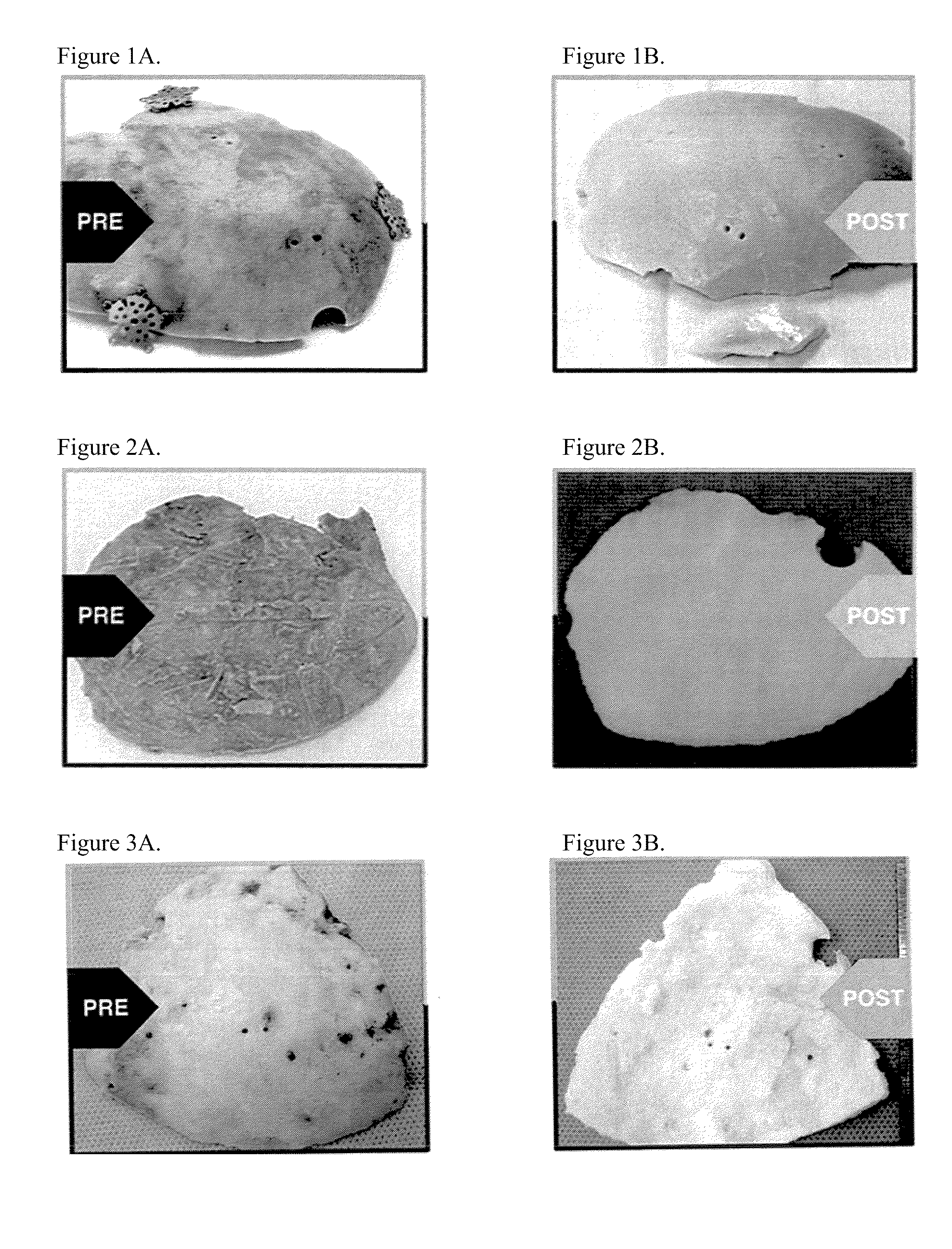
[0074]A right decompressive craniectomy was performed on a 43-year-old patient after a motor vehicle collision. The bone flap removed during the craniectomy was stored in a freezer at −80° C. On the 70th day after the injury, after the patient had recovered, surgeons performed a cranioplasty, using the stored bone flap to close the defect from the previous craniectomy. A week later, the patient presented with clinical symptoms of infection, including running a high fever. The cranial bone flap was removed, the patient was treated for the infection, and the bone flap was sent to LifeNet Health for processing. The bone flap was swabbed for contaminants during surgery and prior to processing at LifeNet Health, and Enterobacter cloacae were cultured from the swabs. (Swab cultures were tested for contaminating agents using both thioglycolate and trypticase soy broth media in a two-temperature / 14-day culture protocol.) The bone flap was then cleaned and disinfected using processes that in...
example 2
[0075]A left decompressive craniectomy was performed on a 20-year-old patient after trauma at Hospital A. The bone flap removed during the craniectomy was stored in a freezer at −80° C. Following his recovery, the patient was transferred to Hospital B for cranioplasty. The bone flap was transported to Hospital B on dry ice, but arrived at room temperature, and it was deemed unsafe for re-implantation. Hospital B stored the bone flap in its freezer at −80° C., until it could be sent to LifeNet Health for processing. At LifeNet Health, the bone flap was swabbed for contaminants, and Staphylococcus epidermis was cultured from the swabs. (Swab cultures were tested for contaminating agents using both thioglycolate and trypticase soy broth media in a two-temperature / 14-day culture protocol.) The bone flap was then cleaned and disinfected using processes that included LifeNet Health's Allowash® technology. Following processing, swab cultures taken from the bone flap were negative. Subseque...
example 3
[0076]A craniectomy was performed during excision of a tumor in an 11-year-old, immune-compromised patient. The patient was immune-compromised due to tumor therapy. The bone flap was sent to LifeNet Health for processing. The bone flap was swabbed for contaminants, and Staphylococcus aureus and Streptococcus viridans were cultured from the swabs. (Swab cultures were tested for contaminating agents using both thioglycolate and trypticase soy broth media in a two-temperature / 14-day culture protocol.) The bone flap was then cleaned and disinfected using processes that included LifeNet Health's Allowash® technology. Following processing, swab cultures taken from the bone flap were negative. Subsequently, the bone flap was treated with a controlled, low dose (i.e., 18 kGy to 58 kGy minimum absorbed dose) of gamma irradiation at low temperatures (i.e., −20° C. to −50° C.). The cleaned and irradiated bone flap was re-implanted in the patient six weeks later. There have been no complication...
PUM
| Property | Measurement | Unit |
|---|---|---|
| temperature | aaaaa | aaaaa |
| temperature | aaaaa | aaaaa |
| temperature | aaaaa | aaaaa |
Abstract
Description
Claims
Application Information
 - R&D
- Intellectual Property
- Life Sciences
- Materials
- Tech Scout
- Unparalleled Data Quality
- Higher Quality Content
- 60% Fewer Hallucinations
Browse by: Latest US Patents, China's latest patents, Technical Efficacy Thesaurus, Application Domain, Technology Topic, Popular Technical Reports.
© 2025 PatSnap. All rights reserved.Legal|Privacy policy|Modern Slavery Act Transparency Statement|Sitemap|About US| Contact US: help@patsnap.com

