Mammography apparatus with X-ray sources arranged at different distances from the chest

a technology of x-ray sources and mammography, which is applied in the direction of instruments, patient positioning for diagnostics, applications, etc., can solve the problems of affecting the evaluation of three-dimensional mammography images, affecting the accuracy of mammography, and reducing the depth resolution of the image, so as to achieve the effect of higher depth resolution

Inactive Publication Date: 2013-12-10
SIEMENS HEALTHCARE GMBH
View PDF9 Cites 4 Cited by
  • Summary
  • Abstract
  • Description
  • Claims
  • Application Information

AI Technical Summary

Benefits of technology

[0009]An object of the present invention is to provide a mammography apparatus and a mammography method that have a higher depth resolution.
[0014]Such a mammography apparatus has the advantage that slice exposures from an additional plane are created for the image reconstruction and volume reconstruction. The three-dimensional reconstruction receives more spatial information. The image reconstruction receives additional data from a plane that is located 90° relative to the chest wall. When a number of x-ray sources or an x-ray source with a number of x-ray positions emitting x-ray radiation are used, additional information is acquired without mechanical movement during a scan in which the x-ray sources are activated in succession and / or x-ray radiation is emitted in succession at different positions of the x-ray source. However, it is also possible to use one or more movable x-ray sources. The positions at which the x-ray radiation is emitted can be located on a horizontal plane across the breast of the patient. The positions can be arranged in a circle, rectangle or square across the breast of the patient. A circular tomosynthesis can thereby be applied. A higher depth resolution and an isotropic resolution of the image plane can be achieved in the circular tomosynthesis. Moreover, the diagnostic data acquisition can take place three-dimensionally, and tissue samples can be examined three-dimensionally.
[0018]The positions emitting the x-ray radiation can be arranged like a matrix. Arbitrary scan trajectories thus can be achieved that can run parallel, orthogonal or at an angle to the shoulder-to-shoulder axis of the patient. The scan trajectories can be freely programmable.
[0019]At a position emitting an x-ray beam that is further removed from the shoulder-to-shoulder axis than those positions that are located nearest to the shoulder-to-shoulder axis, the x-ray radiation can be emitted at an angle relative to vertical in the direction of the breast tissue. The intensity of the x-ray beam striking the x-ray detector thus can achieve a relatively high value without the breast tissue being exposed with a high radiation dose.
[0020]The x-ray source can be a multifocus x-ray tube and / or a microfocus x-ray tube. The x-ray source can generate the x-ray radiation both by means of conventional emitter technology (i.e. thermal emission—and by means of cold emission (i.e. field emitters). The cold emission and the field emitter can be realized by what are known as carbon nanotube emitters, for example. The x-ray source can have only one cathode and one anode, wherein a beam deflection unit has the effect that the x-ray beam is emitted from the desired position. It is also possible to use an x-ray source with one cathode and a plurality of anodes. The x-ray source can have at least one collimator that has the effect that the x-ray radiation passes only through the tissue to be examined and not through other tissue.
[0021]The number of positions at which an x-ray beam can be emitted for an acquisition can correspond to the number of pixels of the x-ray detector. This has the advantage that an image reconstruction is required for a projection image.

Problems solved by technology

These filtered back projection methods present reconstructed images with a high contrast and a high accuracy of detail but lose information about the relative tissue density due to the missing data given tomosynthesis with limited scan angle.
The x-ray attenuation coefficients of these tissue types are very similar, which significantly hinders the evaluation of three-dimensional mammography images.
It is additionally made more difficult in that cancerous tissue has an x-ray attenuation coefficient similar to other tissue types.
The linear scanning over a limited angle range leads to a limited depth resolution.
Therefore, conventional mammography apparatuses and mammography methods have a limited sensitivity and specificity.

Method used


Image

  • Mammography apparatus with X-ray sources arranged at different distances from the chest
  • Mammography apparatus with X-ray sources arranged at different distances from the chest
  • Mammography apparatus with X-ray sources arranged at different distances from the chest

Examples


fourth embodiment

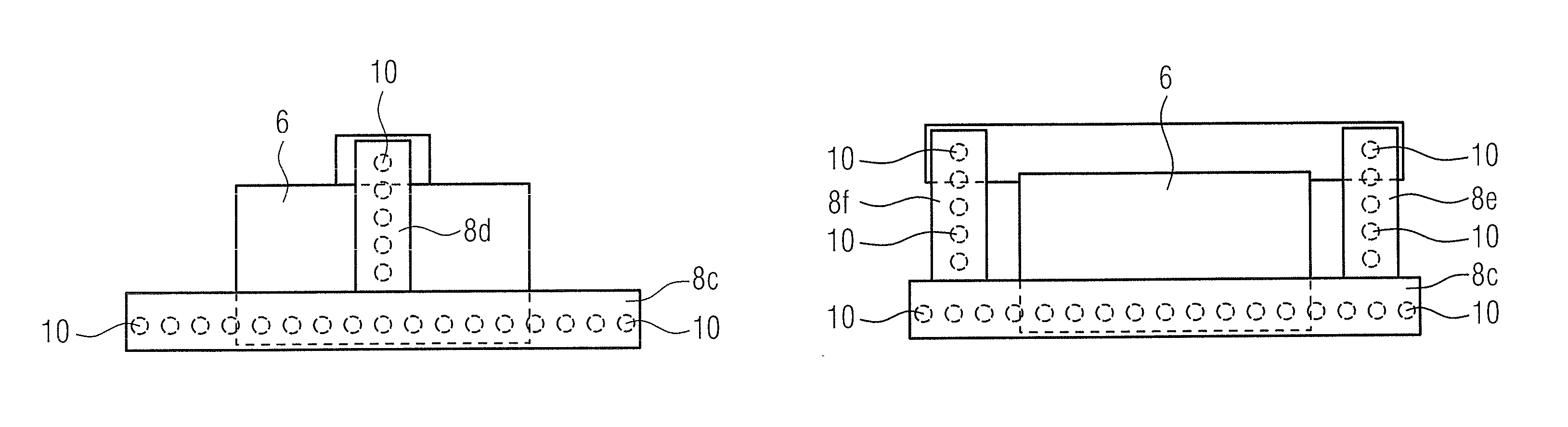
[0049]The fourth embodiment thus has three scan trajectories. The first scan trajectory runs parallel to the shoulder-to-shoulder axis of a patient. The second and third scan trajectory respectively runs orthogonal to the shoulder-to-shoulder axis of the patient. The right multifocus x-ray tube 8e and the left multifocus x-ray tube 8f can be arranged so as to be movable, such that they can be shifted into a position in which optimal acquisition conditions are to be expected. The x-ray beams strike the detector 6 after they have passed through the tissue.

fifth embodiment

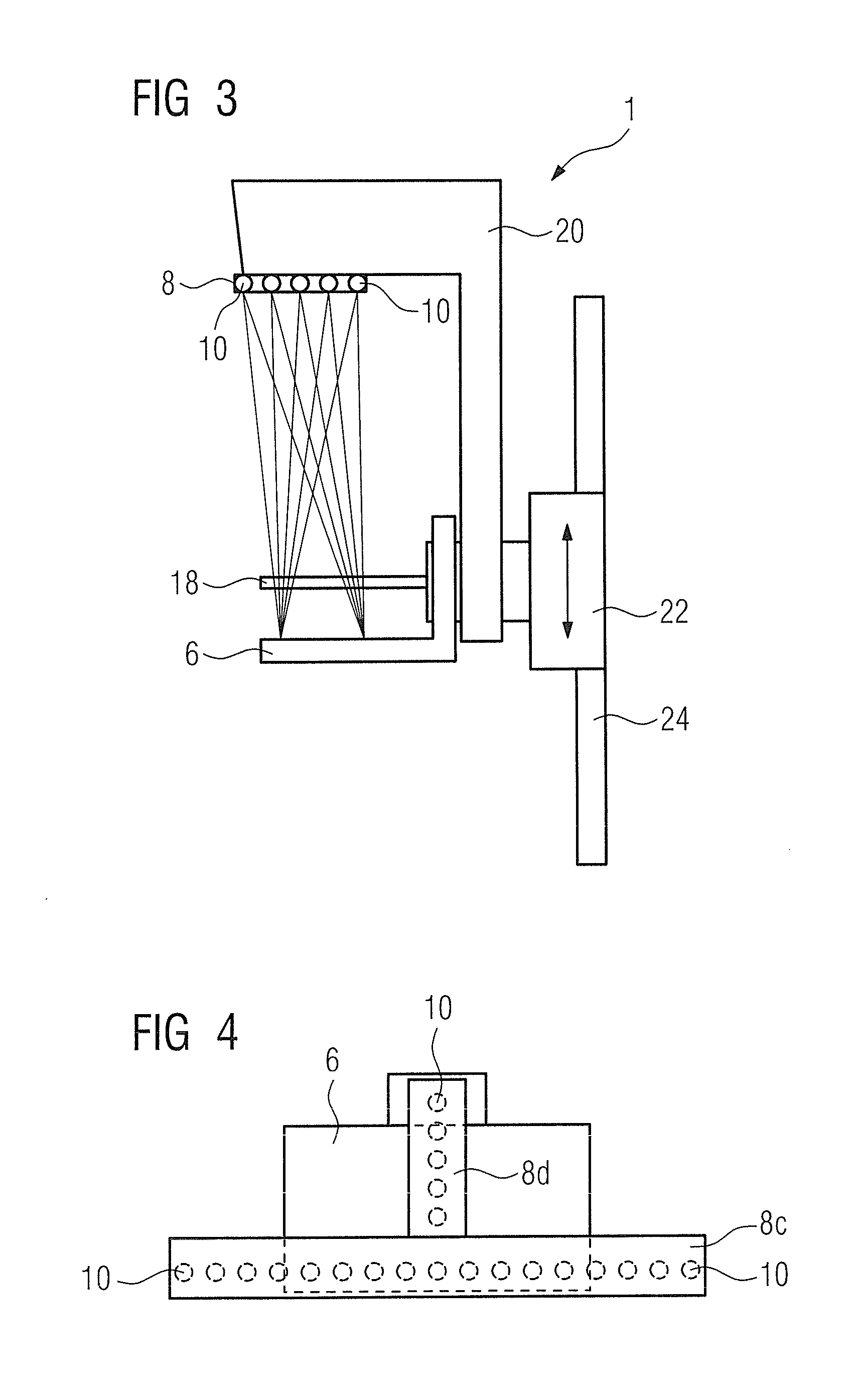
[0050]FIG. 6 shows the invention in which a multifocus matrix x-ray tube 8g is provided. The positions 10 emitting an x-ray beam are arranged in a matrix on the multifocus matrix x-ray tube. A number of scan trajectories are thereby possible that run parallel to the shoulder-to-shoulder axis of a patient. A number of scan trajectories are also possible that run orthogonal to the shoulder-to-shoulder axis of a patient. Moreover, scan trajectories are possible that run at an angle to the shoulder-to-shoulder axis of the patient. The x-ray beams strike the detector 6 after they have passed through the tissue.

[0051]FIG. 7 shows a side view of a position 10 emitting an x-ray beam, the position 10 being located in the second multifocus x-ray tube 8d, the third multifocus x-ray tube 8e, the fourth multifocus x-ray tube 8f or in the multifocus matrix x-ray tube 8g at a position that is further removed from the shoulder-to-shoulder axis of the patient than another position emitting an x-ray ...


PUM


Abstract

A mammography apparatus has at least one x-ray source to emit x-ray radiation and an x-ray detector with a number of pixels. The mammography apparatus is designed so that the x-ray radiation is emitted into the breast tissue of a patient at a number of positions, and the x-ray radiation is detected by the x-ray detector after it has passed through the breast tissue. The positions have different distances from the shoulder-to-shoulder axis of the patient.

Description

BACKGROUND OF THE INVENTION[0001]1. Field of the Invention[0002]The present invention concerns a mammography apparatus in which x-ray sources have different distances from the chest.[0003]2. Description of the Prior Art[0004]In a tomosynthesis procedure, a three-dimensional image is generated from a number of two-dimensional images. A two-dimensional image or a projection of the tissue to be examined that the x-ray beam passes through is generated by an x-ray device with an x-ray source and a detector. The two-dimensional image depicts the attenuation of the tissue in the volume that the beam has passed through. A second two-dimensional image or a second projection of the same tissue or volume is acquired after the radiation source and / or the detector has been moved into a second position. A three-dimensional tomosynthesis image can be generated by a computerized reconstruction after a number of such two-dimensional images have been acquired.[0005]One field of application of the afo...

Claims


Application Information

Patent Timeline
Patent Type & Authority Patents(United States)
IPC IPC(8): A61B6/00
CPCA61B6/025A61B6/4007A61B6/4028A61B6/4233A61B6/502A61B6/4435
Inventor HOERNIG, MATHIASMERTELMEIER, THOMAS
Owner SIEMENS HEALTHCARE GMBH
Why Eureka
Try Eureka