Method and system for imaging a volume using a three-dimensional spiral scan trajectory

a spiral scan and volume technology, applied in the field of medical imaging, can solve the problems of affecting the resolution of the plane parallel, the quality the general limitation of the projection data,

Inactive Publication Date: 2006-05-16
GENERAL ELECTRIC CO
View PDF1 Cites 3 Cited by
  • Summary
  • Abstract
  • Description
  • Claims
  • Application Information

AI Technical Summary

Benefits of technology

[0006]The present technique provides a novel approach to acquiring projection images during tomosynthesis. In particular, the present technique provides for the use of three-dimensional scan trajectories for use in the acquisition of tomosynthesis projection images. The depth resolution of three-dimensional images reconstructed from projection images acquired at different locations along the z-axis may be improved relative to similar images derived from projection data acquired using one and two-dimensional scan trajectories.

Problems solved by technology

For objects that are small relative to the size of the detector, the quality of the projection data, in turn, is generally limited by the range of angles over which the projection data is acquired.
Because depth resolution is limited by the available range of motion of the X-ray source, the depth resolution may not be as good as the resolution in the plane parallel to the detector surface.

Method used


Image

  • Method and system for imaging a volume using a three-dimensional spiral scan trajectory
  • Method and system for imaging a volume using a three-dimensional spiral scan trajectory
  • Method and system for imaging a volume using a three-dimensional spiral scan trajectory

Examples


Embodiment Construction

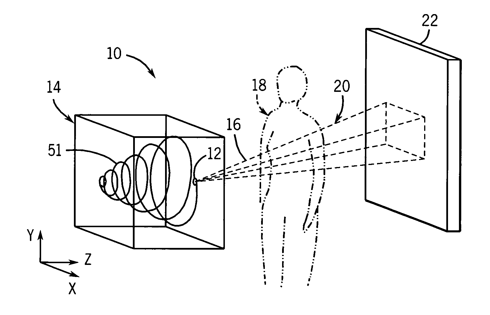
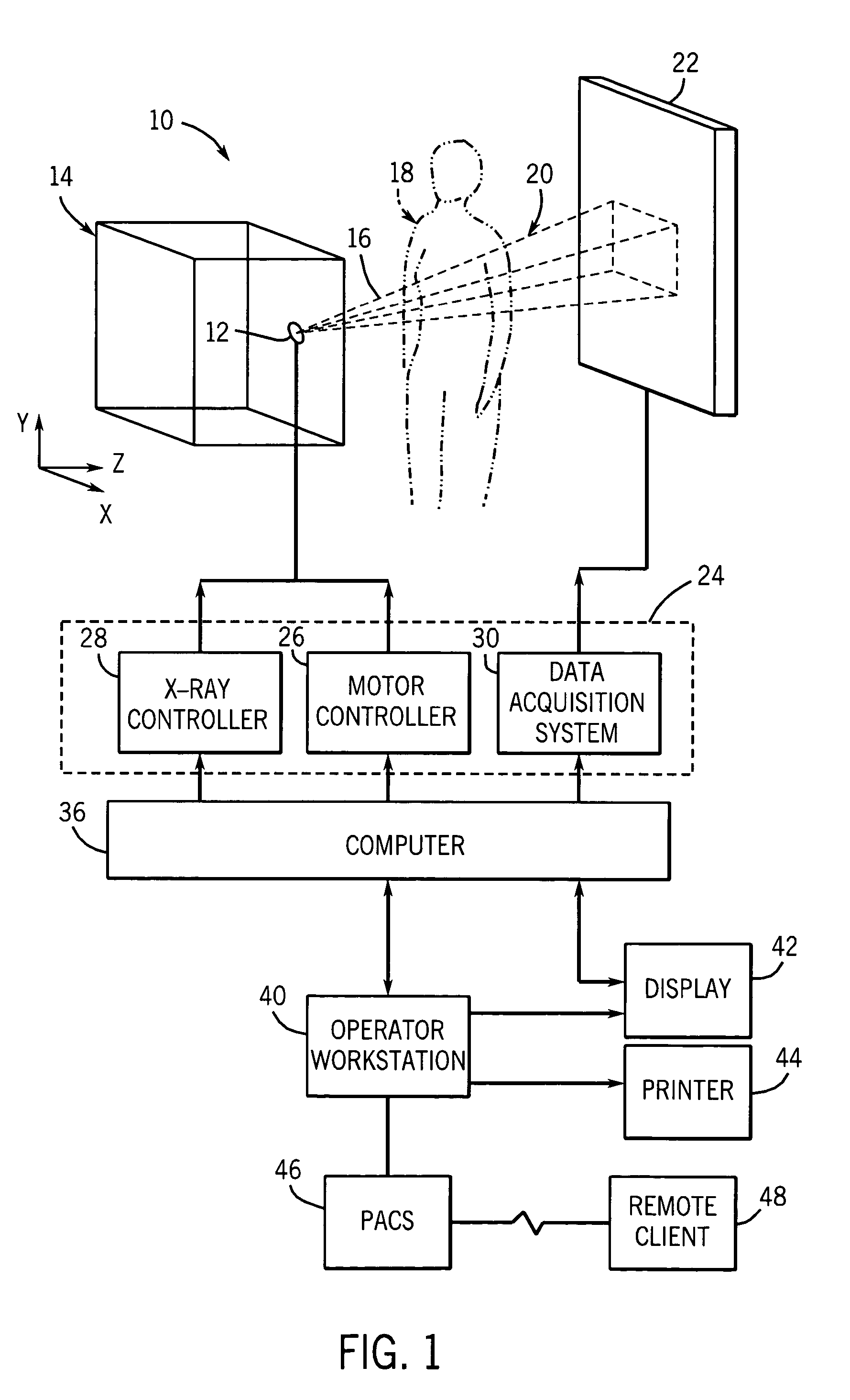
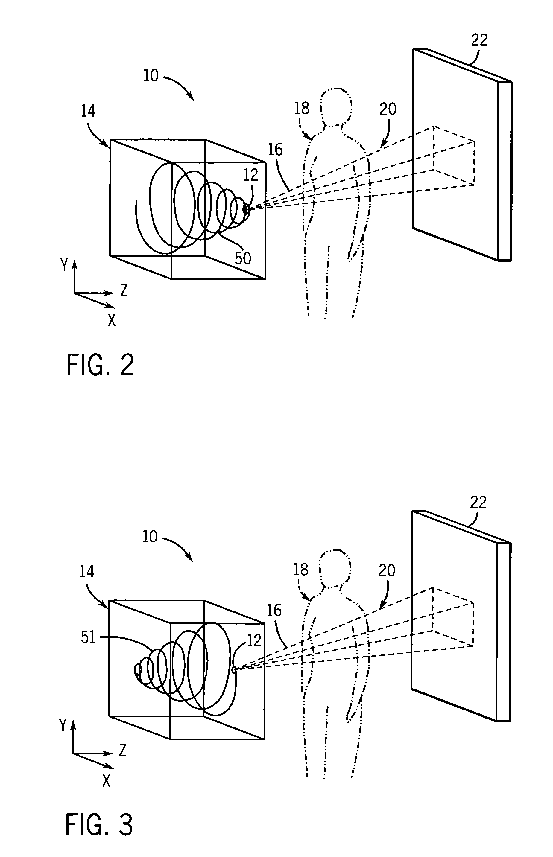
[0016]In the field of medical imaging, various imaging modalities may be employed to non-invasively examine and / or diagnose internal structures of a patient using various physical properties. One such modality is tomosynthesis imaging which utilizes a limited number of projection radiographs that are each acquired at a different angle relative to a patient. The projection radiographs may be combined to generate a set of data that provides three-dimensional context and structure for the volume of interest. Typically, the projection radiographs are generated using an X-ray source moving in a plane parallel to a detector. The X-ray source may move in one or two dimensions within the plane. The linear and / or planar movement of the X-ray source effectively limits the depth resolution that may be achieved in three-dimensional images reconstructed from the acquired projection data. The present technique is directed to the improvement of depth resolution by incorporating motion along the de...


PUM

PropertyMeasurementUnit
heightsaaaaaaaaaa
depthaaaaaaaaaa
tomosynthesis imagingaaaaaaaaaa

Abstract

A technique is provided for improving the depth resolution of tomosynthesis images. The technique provides for the use of spiral scan trajectories. The X-ray source may moved along the spiral scan trajectory, acquiring projection data at various locations on the trajectory which are offset in the z-direction, i.e., in a direction perpendicular to the detector. Projection data acquired at different positions in the z-direction may be used to generate three-dimensional, tomosynthesis images having improved depth resolution.

Description

BACKGROUND OF THE INVENTION[0001]The present invention relates generally to the field of medical imaging, and more specifically to the field of tomosynthesis. In particular, the present invention relates to the use of three-dimensional scan trajectories during acquisition of image data.[0002]Tomosynthesis is an imaging modality that may be used, in a medical context, to allow physicians and radiologists to non-invasively obtain three-dimensional representations of selected organs or tissues of a patient. In tomosynthesis, projection radiographs, conventionally known as X-ray images, are acquired at different angles relative to the patient. Typically, a limited number of projection radiographs are acquired over a relatively small angular range. The projections comprising the radiographs generally represent the line integrals of the attenuation coefficients along the respective X-ray paths through the patient and, therefore, convey useful data regarding internal structures.[0003]From ...

Claims


Application Information

Patent Timeline
Patent Type & Authority Patents(United States)
IPC IPC(8): A61B6/03A61B6/00A61B6/04G01N23/00G21K1/12H05G1/60
CPCA61B6/025A61B6/4021A61B6/4233A61B6/466A61B6/027A61B6/4429Y10S378/901
Inventor AL-KHALIDY, ABDULRAHMANEBERHARD, JEFFREY WAYNEALYASSIN, ABDALMAJEID
Owner GENERAL ELECTRIC CO
Why Eureka
Try Eureka