[0010]The first patient-specific jig can be a hemispherical shaped device or a sub-hemispherical shaped device, and can be comprised of composite material or other materials. The first patient-specific jig may include at least three alignment members for attachment to specific portions of the jig based on the specific bone structure of the patient's coxal bone. The at least three alignment members may assist with proper alignment of the first patient-specific jig in the patient's acetabulum during surgery. The three alignment members may include first, second, and third alignment members that are positioned at specific outer portions of the first patient-specific jig.
[0011]In some embodiments, the first, second, and third alignment members are designed and adapted to be hooked on or engaged with (or otherwise positioned at) particular portions of the coxal bone adjacent to the acetabulum to stabilize and properly orient the first patient-specific jig into the acetabulum during surgery. The first alignment member may be positioned on the first patient-specific jig to engage a particular portion of the medial rim of the acetabulum of the coxal bone. The second alignment member may be positioned on the first patient-specific jig to engage a particular portion of the greater sciatic notch of the coxal bone. The third alignment member may be positioned on the first patient-specific jig to engage a particular portion of the obturator foramen of the coxal bone. These alignment members may contact any three areas of bone peripheral to the acetabulum. Thus, the first patient-specific jig includes three reference points / members, specific to the patient's acetabulum of the coxal bone, to properly align the first patient-specific jig in the acetabulum during surgery and provide for proper orientation of the reaming machine when resurfacing the acetabulum.
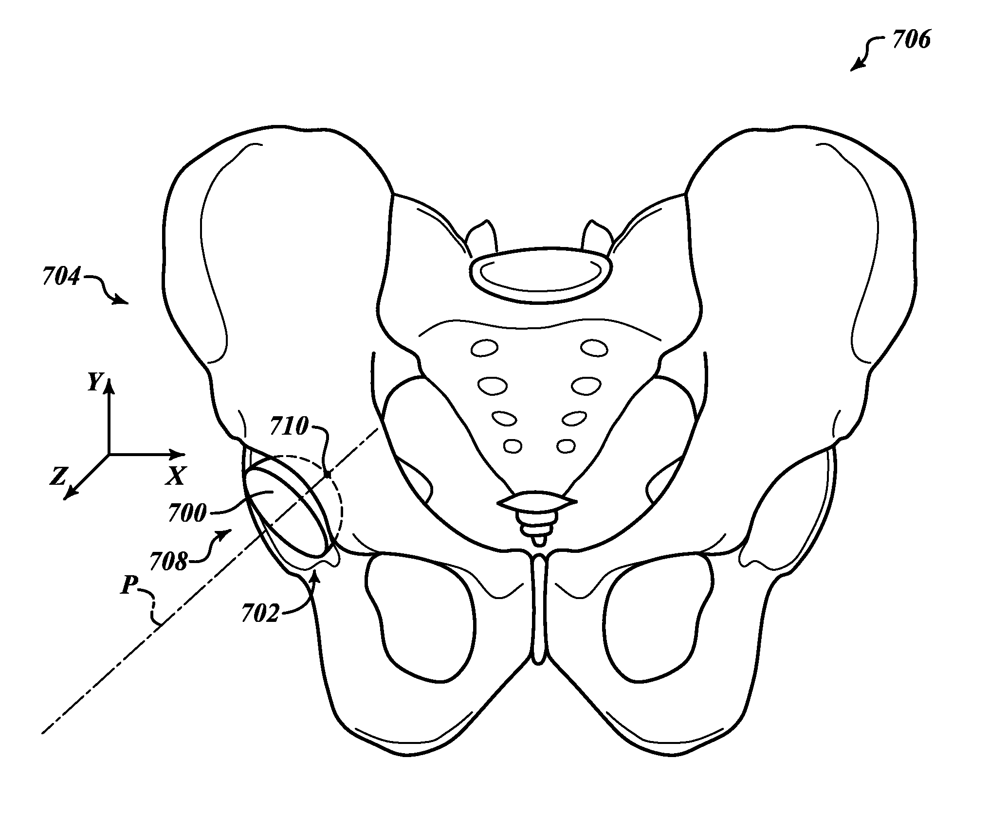
[0013]An aperture may be provided radially into or through the first patient-specific jig. The aperture is adapted to receive a guide pin or post extension through the aperture during surgery. The guide pin or extension is placed into the aperture and is removably secured to the coxal bone of the patient in a particular orientation and at a particular depth, as determined by the surgeon during preoperation. The first patient-specific jig is then removed while the guide post remains positioned in the coxal bone at the desired angle and position. The guide post may then serve as a guide for the reaming machine to accurately ream (resurface) the acetabulum to a predetermined depth and orientation for receiving the acetabular cup. The guide post and the reamer may have the same or similar central axes, as with conventional reaming machines and processes. In some embodiments the guide post may be removed prior to reaming and a surgeon at his discretion may use the guide post sinus tract as a guide to reaming without the guide post at a desired angle and to a desired depth.
[0014]As discussed above, it is critical to ream the acetabulum accurately and as determined during preoperation. Accordingly, the second patient-specific jig may be provided to assist in determining accurate reaming and proper alignment of the acetabular cup before the cup is implanted into the patient's acetabulum. As indicated below, a third patient-specific jig may also be used to progressively prepare the acetabulum to properly receive the prosthetic acetabular cup.
[0019]The predetermined orientation of the apertures and the alignment members of both the first and second (and, as applicable, third) patient-specific jigs may provide the surgeon with a quick, accurate means to properly resurface the acetabulum without the use of additional devices and machines. This is possible because the positions of the alignment members of the first, second and third patient-specific jigs are based upon the patient's bone structure, thereby providing three reference points to accurately utilize the first jig, the guide post, the reamer, and the second and third jigs, as planned during preoperation based upon the 3D modeling images of the patient.

