Device and method for bone imaging

a bone imaging and device technology, applied in the field of medical devices, can solve the problems of insufficient accuracy of 5 degrees for the determination of fusion, difficult assessment of healing, and insufficient resolution of diagnostic techniques such as mri or ct to determine if healing has occurred

Inactive Publication Date: 2015-06-09
HALIFAX BIOMEDICAL
View PDF13 Cites 3 Cited by
  • Summary
  • Abstract
  • Description
  • Claims
  • Application Information

AI Technical Summary

Benefits of technology

[0022]wherein the less the difference in position of the implant in the unloaded state as compared to the load-bearing state, the better the assessment.

Problems solved by technology

However, assessment of healing is difficult.
Diagnostic techniques such as MRI or CT do not have sufficient resolution to determine if healing has occurred.
Flexion extension comparisons using MRI have reported accuracy of 5 degrees which is not sufficient for the determination of fusion.

Method used


Image

  • Device and method for bone imaging
  • Device and method for bone imaging

Examples


Embodiment Construction

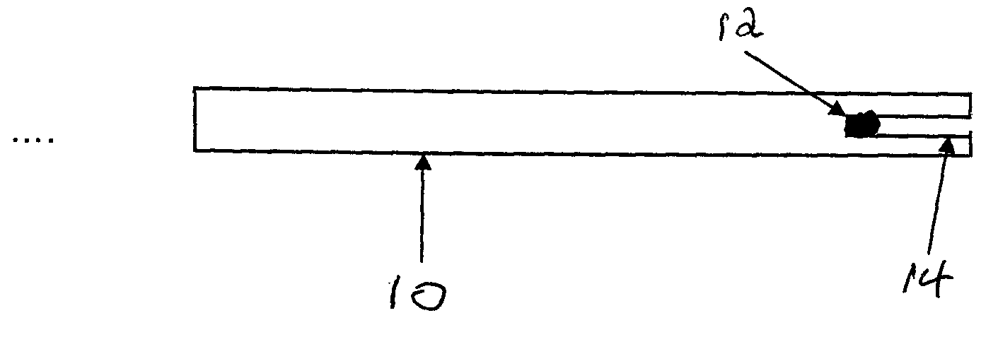
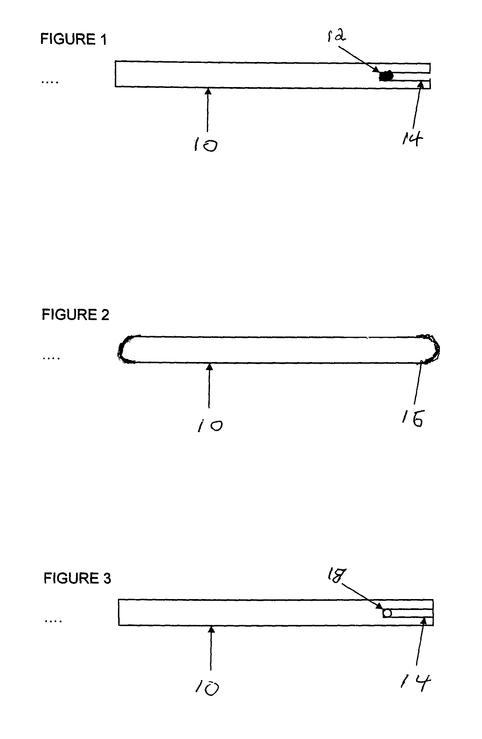
[0028]The invention relates to modified medical implants that can be used in bone assessment. The modified implant includes an implant modified with a detectable marker that can readily be detected to determine the position of the modified implant.

[0029]The term “bone assessment” is used herein to encompass the assessment of bone healing following injury by fracture or surgical intervention that create the same biological conditions as an injury, assessment of surgical fusions such as spine and ankle fusion operations, the assessment of bone growth and bone motion for any other reason.

[0030]In one aspect of the invention, medical implants comprising small radio-opaque elements as the detectable marker are provided in which these elements are embedded or attached to the medical implant. These elements are more radio-opaque than the medical implant itself. The result is that in an x-ray of the implant / bone construct, the embedded elements can be clearly seen. The embedded elements are...


PUM

PropertyMeasurementUnit
medical imagingaaaaaaaaaa
distanceaaaaaaaaaa
rigidityaaaaaaaaaa

Abstract

The invention comprises a modified implant useful to assess bone by imaging. The modified implant comprises a medical implant and at least one detectable marker element associated with the implant that serves as a reference point in medical imaging. Preferably, the implant and the detector marker elements have different radiolucencies such that one element can be seen through the other in medical imaging.

Description

FIELD OF INVENTION[0001]The present invention relates to medical devices, and in particular, a medical implant that is useful in bone imaging to track bone growth, healing and motion.BACKGROUND OF THE INVENTION[0002]Medical implants are frequently used to support bones during healing. However, assessment of healing is difficult. Diagnostic techniques such as MRI or CT do not have sufficient resolution to determine if healing has occurred.[0003]The current standard for the assessment of fracture healing is a planar radiograph and the current standard for assessment of spinal fusion for example is direct palpation of the spine in a second exploratory operation. Flexion extension comparisons using MRI have reported accuracy of 5 degrees which is not sufficient for the determination of fusion.[0004]Thus, there exists a need to establish a method and device that provides assessment of fusion and bone healing. Having the ability to assess bone healing would assist surgeons in clinical dec...

Claims


Application Information

Patent Timeline
Patent Type & Authority Patents(United States)
IPC IPC(8): A61B5/103A61B6/12A61B19/00A61L31/18
CPCA61L31/18A61B6/12A61B19/54A61B2019/5416A61F2250/0032A61F2250/0098A61L2430/02A61B2090/3916A61B90/39
Inventor MUNRO, CHAD
Owner HALIFAX BIOMEDICAL
Why Eureka
Try Eureka