Pre- and intra-operative imaging of bladder cancer
a bladder cancer and intraoperative imaging technology, applied in the field of pre- and intra-operative imaging of bladder cancer, can solve the problems of high false-negative rate of methods, high spatial resolution, and difficult to accurately perform biopsies in patients
- Summary
- Abstract
- Description
- Claims
- Application Information
AI Technical Summary
Problems solved by technology
Method used
Image
Examples
example 1
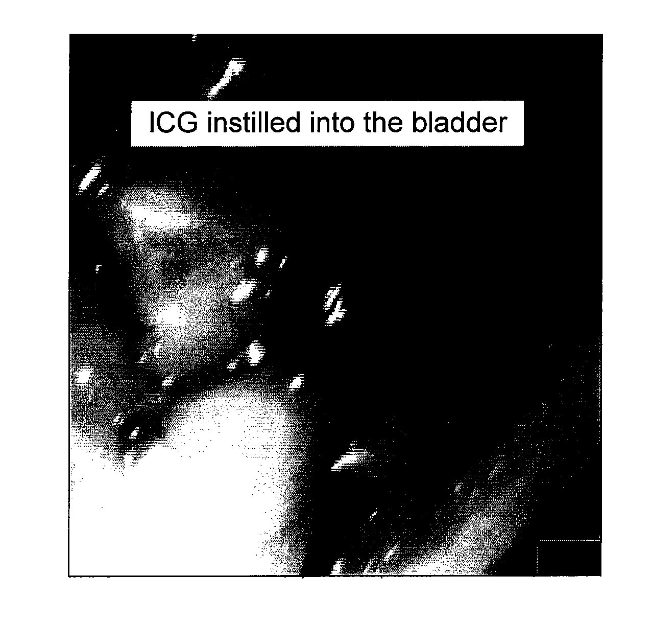
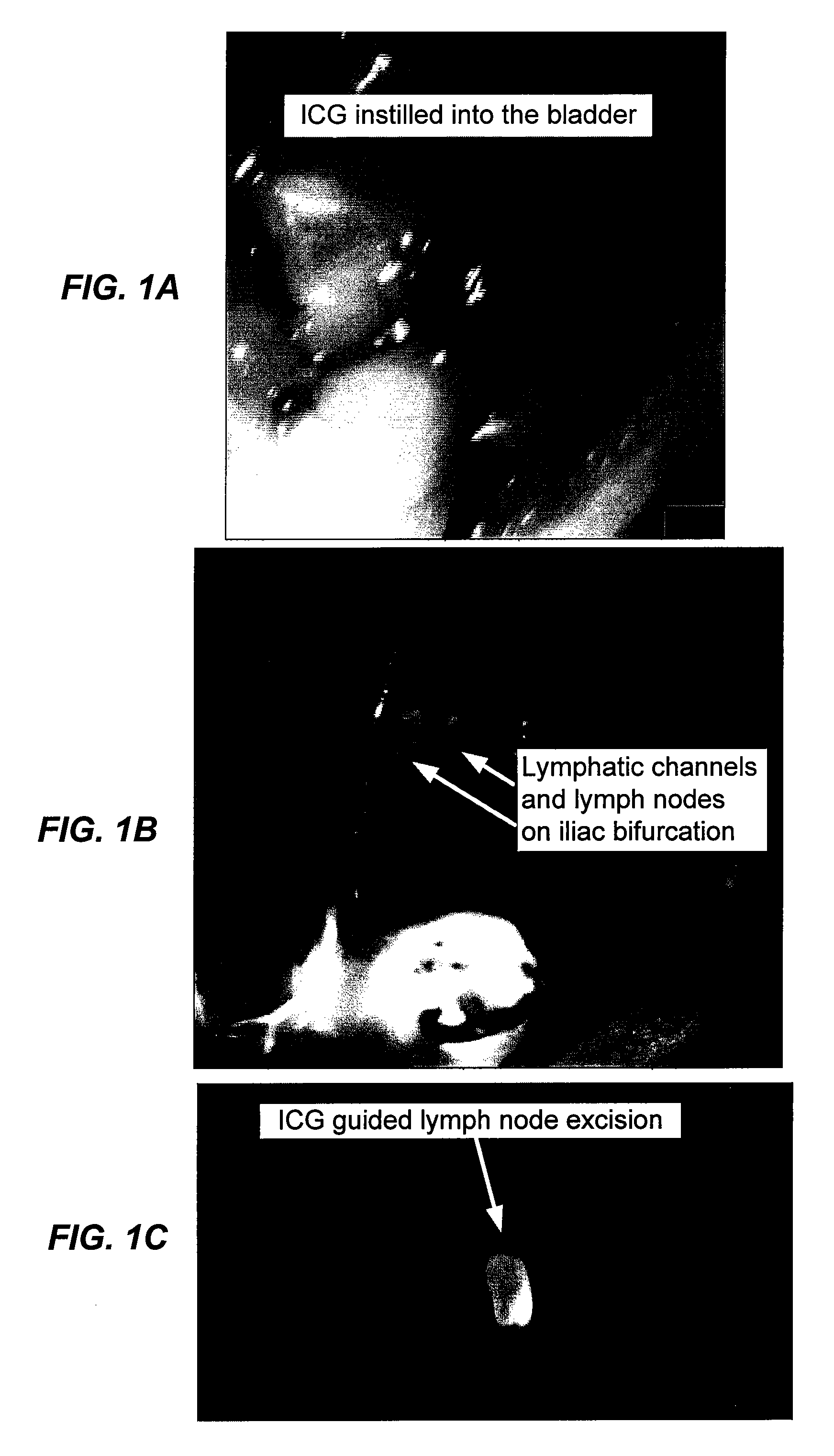
[0039]UPII-SV40T transgenic mice were used that express the SV40 T antigen specifically in the urothelium and that reliably develop bladder tumors. Twelve female mice received intravesical instillations of ICG (2.5 mg / mf, 100 or 200 μl) and 20, 30 and 60 minutes later underwent intraoperative near infrared fluorescence (“NIRF”) identification of lymphatic channels and SLNs. Wildtype female mice were controls. NIRF and H&E microscopy were used to confirm presence of the lymphatic tissue and eventual metastasis.
[0040]All 12 animals receiving ICG showed draining lymphatic channels and SLNs. At least two sentinel nodes were detected in all animals. SLNs were in common or external iliac lymphatic chains. In one mouse, SLNs were detected in presacral and paravesical chain. SLNs were not visible in normal saline control group. IRF and H&E microscopy confirmed presence of lymphatic tissue in resected SLNs.
PUM
 Abstract
Description
Claims
Application Information
 - R&D
- Intellectual Property
- Life Sciences
- Materials
- Tech Scout
- Unparalleled Data Quality
- Higher Quality Content
- 60% Fewer Hallucinations
Browse by: Latest US Patents, China's latest patents, Technical Efficacy Thesaurus, Application Domain, Technology Topic, Popular Technical Reports.
© 2025 PatSnap. All rights reserved.Legal|Privacy policy|Modern Slavery Act Transparency Statement|Sitemap|About US| Contact US: help@patsnap.com


