Method and system for quantitative renal assessment

a quantitative and renal assessment technology, applied in the field of medical imaging analysis, can solve the problems of limiting the application of models, requiring physical display of images, laborious interpretation,

Active Publication Date: 2015-11-03
CHILDRENS HOSPITAL OF LOS ANGELES
View PDF9 Cites 6 Cited by
  • Summary
  • Abstract
  • Description
  • Claims
  • Application Information

AI Technical Summary

Problems solved by technology

These techniques were entirely manual, requiring physical display of the images and laborious interpretation by the diagnostician.
However, the complexity of renal physiology and variance in imaging techniques sometimes complicates the implementation of a sophisticated model approaching a realistic kidney, which limits the application of those models.

Method used


Image

  • Method and system for quantitative renal assessment
  • Method and system for quantitative renal assessment
  • Method and system for quantitative renal assessment

Examples


example 1

Experimental Methods of the Invention
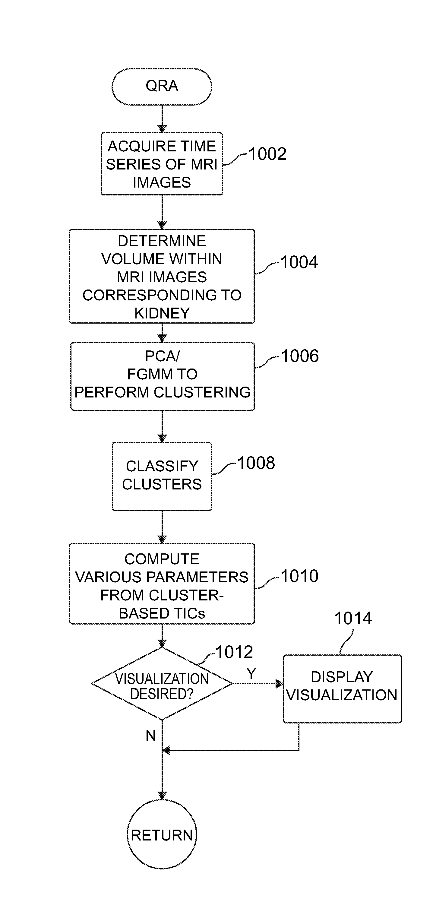
[0086]MRI Protocol

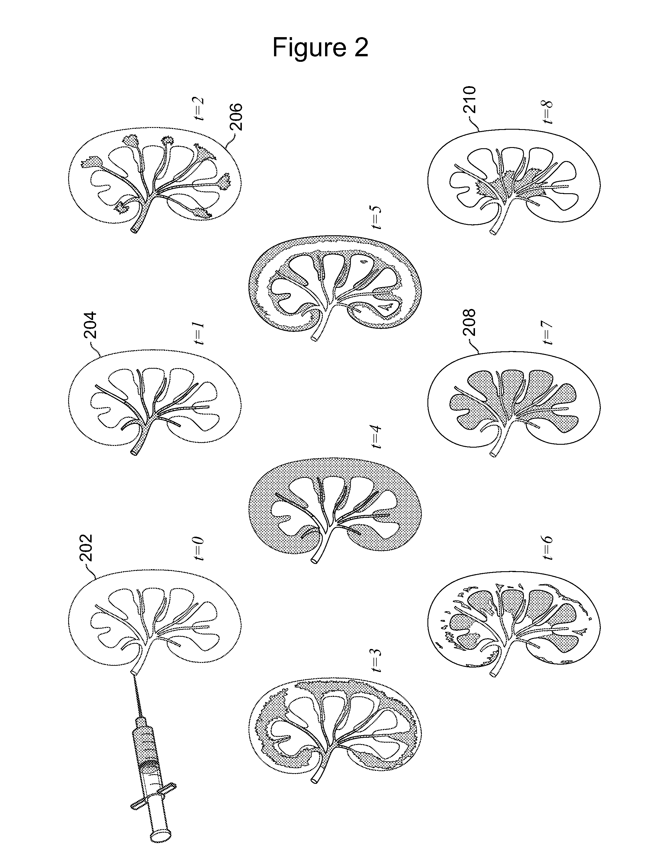
[0087]A dynamic MRI with 3D coronal fast GRE sequence was performed on a 1.5 Tesla system. For kidney diagnosis, coils are put near the abdomen area. After a bolus of contrast (Gd-DTPA) was injected, data were acquired with each set of images taking about 30 seconds and the total procedure lasting about 20 minutes. The initial matrix size was 256*256, flip angle is 45, TR range is from 6 to 6.9 ms, and TE range is from 0.83 to 1.27 ms. To keep the linear relationship between the image intensity and the contrast concentration, a dose of the contrast is injected 0.2 mg / kg.

[0088]Preprocessing

[0089]The target of the preprocessing step is to define an interesting kidney region and de-noise the MRI data. To focus on the relevant area, the user selects a rectangular region that includes the kidney in a maximum intensity projection (MIP) image. To decrease the noise influence, a bilateral filter is adopted for each volume of data in spa...

example 2

Results

[0130]Digital Phantom

[0131]To test the capacity of dealing with noise, a 4D digital phantom is designed for the experiments herein, which is spatially composed of three inner compartments, and has time series activities in the fourth (temporal) dimension. The average time series activities are calculated from real data and used as typical TICs for each compartment.

[0132]Fifty trials were performed by adding Gaussian white noise, with variance increasing from 10−5 to 1, to the digital phantom. Simultaneously, the signal to noise ratio (SNR) decreased from 56.5 to 0.7. The SNR is defined as ratio of the average signal and RMS (root mean square) noise in the early enhanced phase.

[0133]The inventors compare the segmented results of the method (R1) with the ground truth (R2) for kidney masks as well as inner compartments. The overlap percent (OP) is defined as Equ. 1 for the similarity measurement and the results are displayed in FIG. 17.

[0134]O⁢⁢P=12⁢(R1⁢⁢R2R1+R1⁢⁢R2R2)(1)

[0135]F...

example 3

Discussion

[0147]The inventors have demonstrated a processing workflow based on clusters of dynamic kidney MRI data. The clusters group voxels with similar TICs in a 3D manner and supply a more precise visual description to allow faster clinical insight. Based on the clusters, automatic methods for kidney masking and compartmental identification are also designed, which is important for both anatomical and functional analysis. To provide a more intuitive visualization for quantification parameters, multiple options and interactions are supplied.

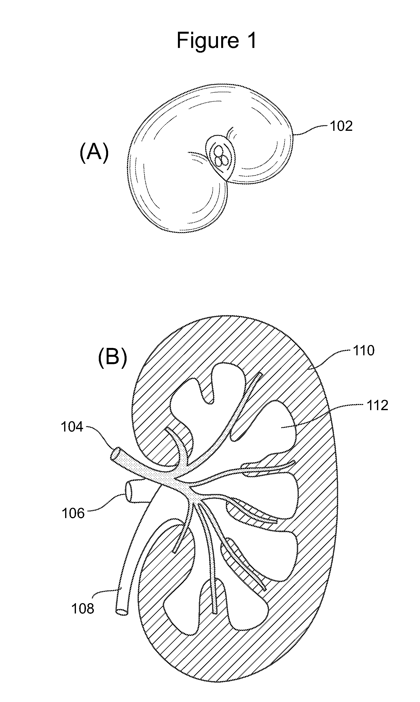
[0148]In the inventors' method some assumptions are made during the processing. First, for the inner compartments, three tissues are taken into account including cortex, medulla and collection system. Although there are more than three compartments in the state-of-the-art kidney model, for the current imaging protocol only these three partitions were practical in the instant study. For example, the more advanced multi-compartment model include...


PUM


Abstract

Method and system embodiments of the present invention employ computational analysis tools running on one or more computer systems to quantitatively analyze MRI data from patients in order to both assess various functional parameters and characteristics of a patient's kidney, as well as to provide an electronic display device interconnected with the one or more computer systems, and accessible visualization of the quantitative assessment to facilitate diagnosis of various types of kidney abnormalities and pathologies.

Description

CROSS-REFERENCE TO RELATED APPLICATIONS[0001]This application is the National Phase of International Application PCT / US2011 / 36797, filed May 17, 2011, which designated the U.S. and that International Application was published under PCT Article 21(2) in English. The present application also claims the benefit of the filing date of U.S. Provisional Application No. 61 / 345,558, filed May 17, 2010, the disclosures of each of which are incorporated herein by reference in their entirety.FIELD OF INVENTION[0002]The present invention is related to medical-imaging analysis and, in particular, to an automated and / or semi-automated method for quantitative assessment of renal function and morphology from a time series of magnetic-resonance images (“MRIs”) of a kidney. The invention furthers the practice of evidence based medicine by providing objective analysis of the normal abnormal and irrelevant.BACKGROUND OF THE INVENTION[0003]All publications herein are incorporated by reference to the same...

Claims


Application Information

Patent Timeline
Patent Type & Authority Patents(United States)
IPC IPC(8): A61B5/05G06T7/00A61K49/06A61M5/00A61B5/055
CPCA61B5/055A61K49/06A61M5/007G06T7/0012G06T2207/10088G06T2207/30084
Inventor MOATS, REXTANG, YANGSHARMA, PRIYANKJACKSON, HOLLIE
Owner CHILDRENS HOSPITAL OF LOS ANGELES
Why Eureka
Try Eureka