Systems and methods for interactive image analysis of digital pathology images

Active Publication Date: 2017-03-07
FLAGSHIP BIOSCI
View PDF5 Cites 1 Cited by
  • Summary
  • Abstract
  • Description
  • Claims
  • Application Information

AI Technical Summary

Benefits of technology

[0008]Data processing systems and methods are provided that improve the perceived efficiency of image analysis tools in a digital pathology workflow when challenged by analyzing large size microscopic images. Various embodiments utilize a separation of the image analysis programs into a first automatically executed segment that requires no user interactions for identification and quantific

Problems solved by technology

The process of running image analysis programs, such as those for pathology analysis, on these large data images, tends to consume a considerable amount of time.
Depending on the sophistication of the programs, analyzing an image of a whole slide tissue section can take up to seve

Method used


Image

  • Systems and methods for interactive image analysis of digital pathology images
  • Systems and methods for interactive image analysis of digital pathology images
  • Systems and methods for interactive image analysis of digital pathology images

Examples


Example

EXAMPLE

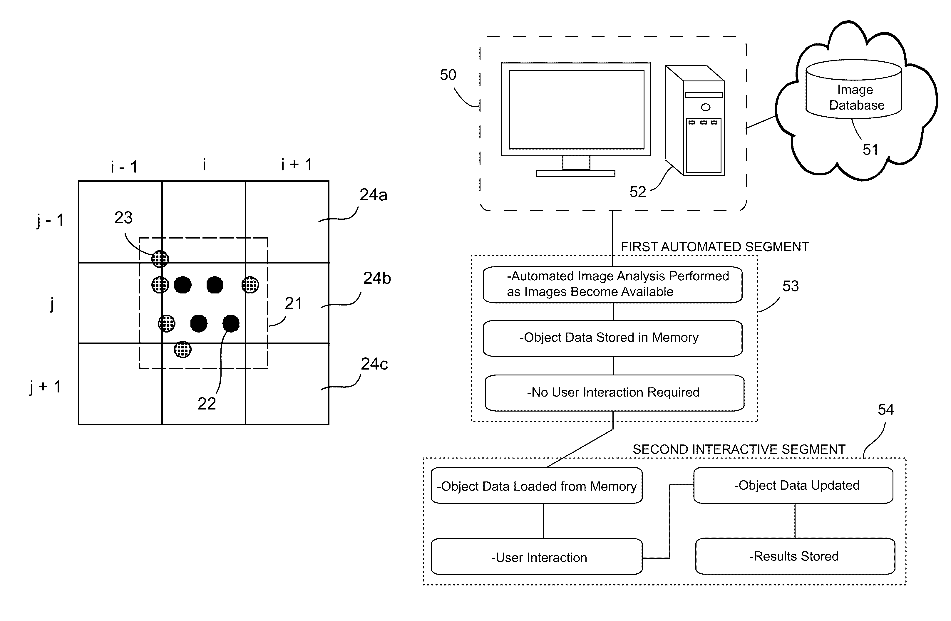
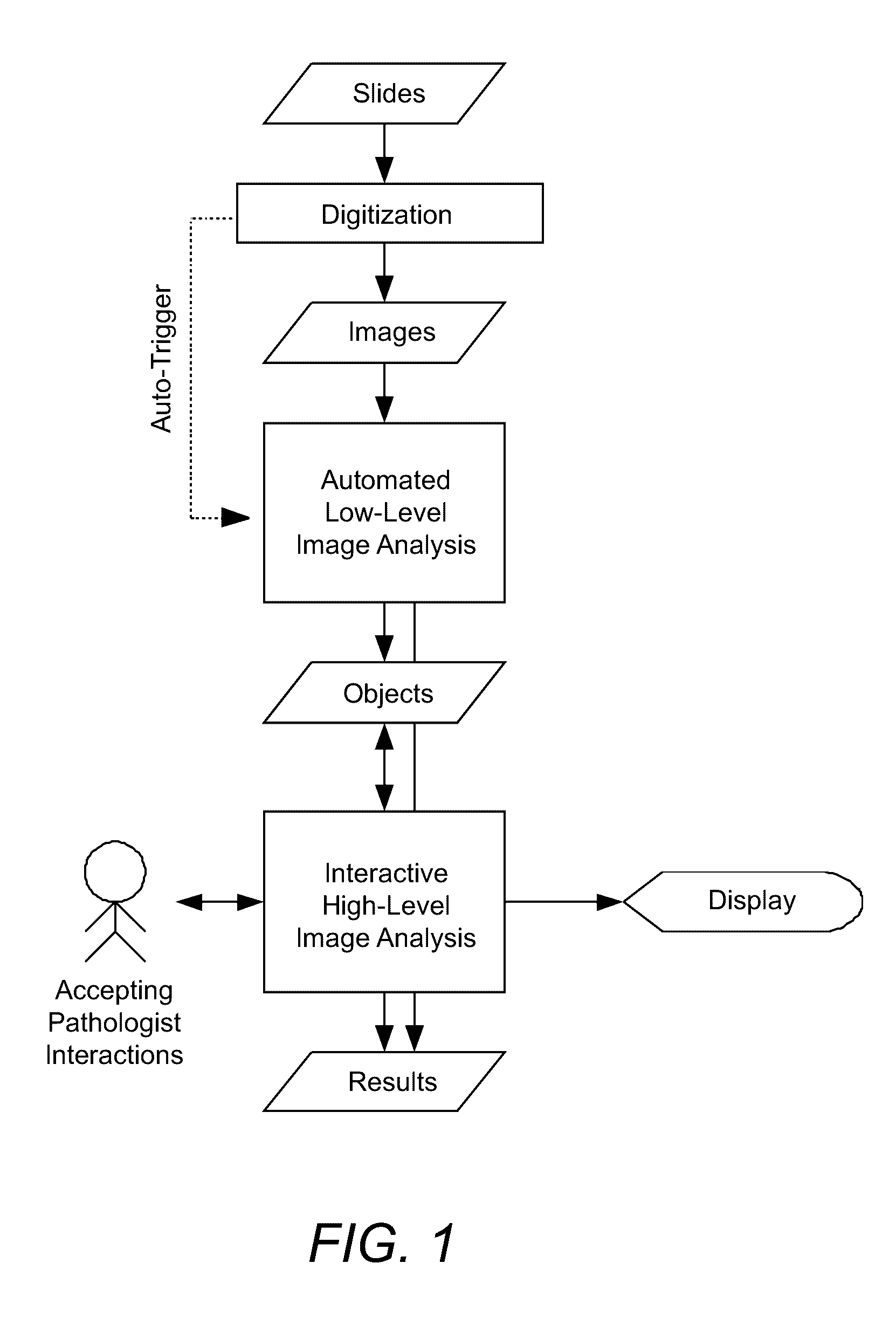
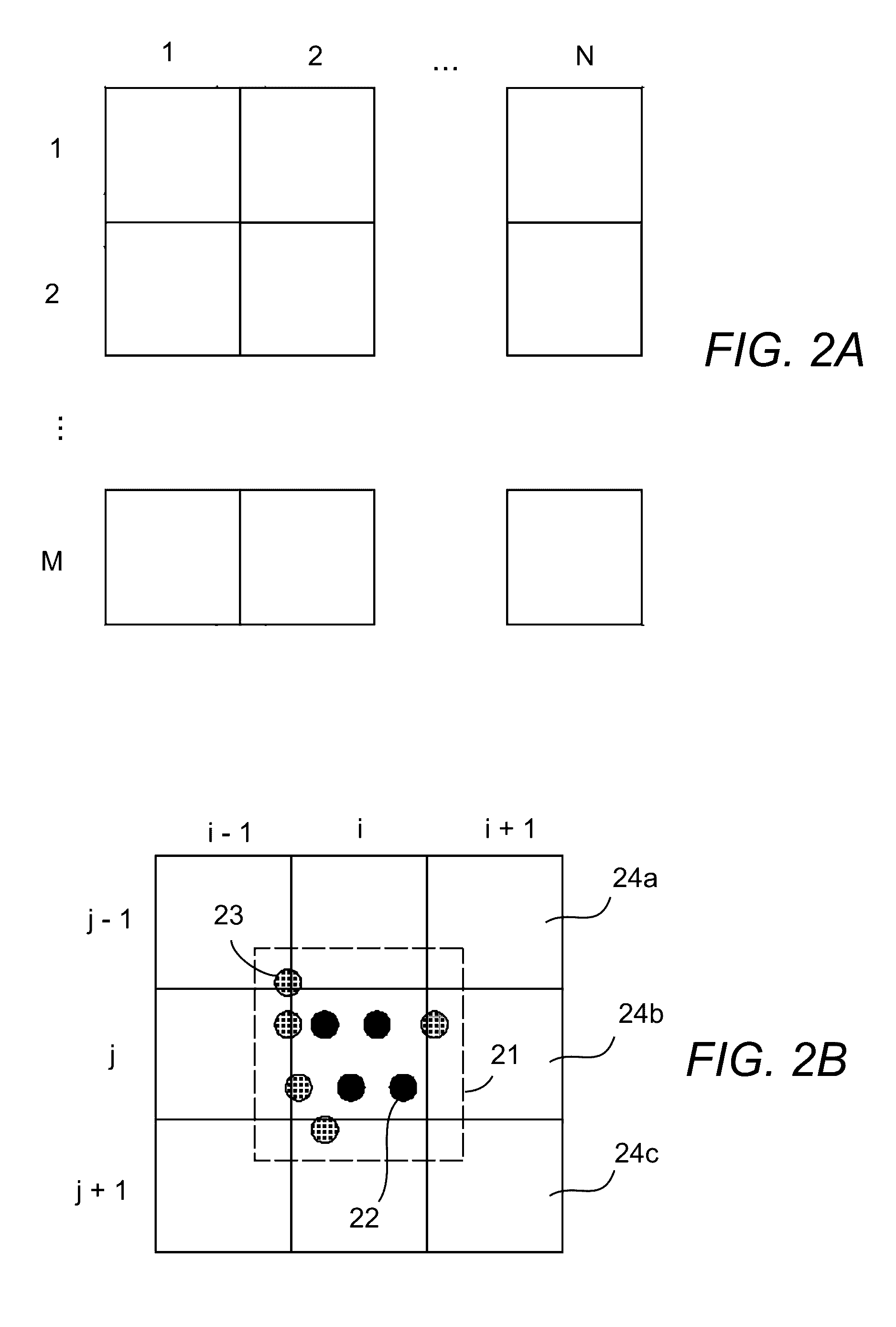
[0050]For illustrative purposes, one example may include the quantitative analysis of progesterone receptors in breast tissue. The image analysis task that needs to be performed consists of detecting the nuclei of invasive tumor cells, measuring the mean optical density of the progesterone preceptor staining on the nuclei and then classifying the nuclei into 4 categories (0—negative, 1—low positive, 2—medium positive, and 3—high positive) based on the amount of progesterone preceptor staining. From the percentages of nuclei in the different categories an H-score is calculated, which essentially is the average category of the nuclei times 100. The program is not able to detect the invasive tumor cells automatically, therefore a user needs to determine which of the cells are invasive tumor cells.

[0051]The automated low-level image analysis program detects all nuclei on the entire slide, quantifies the progesterone preceptor staining and provides the classification of the nuclei...


PUM


Abstract

Data processing systems and methods are provided that improve the perceived efficiency of image analysis tools in a digital pathology workflow when challenged by analyzing large size microscopic images. Various embodiments utilize a separation of the image analysis programs into a first automatically executed segment that requires no user interactions for identification and quantification of object data of processed images, and a second interactive segment for accepting user input. User interactions are only applied to the already calculated object data resulting in an improved system response time. A tile-based processing scheme with concurrent processing and generation of object data in a tile-based data format provides further performance improvements of the system.

Description

CROSS-REFERENCE TO RELATED APPLICATIONS[0001]This application claims benefit of priority with U.S. Provisional Application Ser. No. 61 / 769,068, filed Feb. 25, 2013.BACKGROUND[0002]Field of the Invention[0003]This invention relates to digital pathology image analysis tools.[0004]Description of the Related Art[0005]Microscopes provide high-resolution imagery that, when digitized, often provide very large digital images. For example, typical whole slide tissue sections acquired with a 20× or 40× objective often yield image sizes of 25 k×25 k pixels or 100 k×100 k pixels, respectively. The process of running image analysis programs, such as those for pathology analysis, on these large data images, tends to consume a considerable amount of time. Depending on the sophistication of the programs, analyzing an image of a whole slide tissue section can take up to several hours.[0006]Where a user is required to run image analysis programs, this results in a complex workflow. The user must firs...

Claims


Application Information

Patent Timeline
IPC IPC(8): G06K9/00
CPCG06K9/00127G06V20/69G06V10/945G06F18/40
Inventor LANGE, HOLGERJOHNSON, TREVOR
Owner FLAGSHIP BIOSCI
Why Eureka
Try Eureka