Medical device for reconstruction of a humerus for the operative treatment of a proximal humerus fracture

a humerus and humerus technology, applied in the field of humerus reconstruction for the operative treatment of a proximal humerus fracture, can solve the problems of inconvenient treatment, inability to grow together or not, permanent stiffness, etc., and achieve the effect of efficient treatmen

Active Publication Date: 2017-04-11
IMPLANT SERVICE BV
View PDF33 Cites 12 Cited by
  • Summary
  • Abstract
  • Description
  • Claims
  • Application Information

AI Technical Summary

Benefits of technology

[0009]An aim of the present invention is to provide an implantable medical device for reconstruction of a humerus for treatment of a proximal humerus fracture, which provides flexibility during the reconstruction of the humerus of a patient. This flexibility makes a more efficient treatment of a proximal fracture of the humerus possible, in particular when the humerus is broken in three or more bone fragments.

Problems solved by technology

If the bone fragments are dislocated, the position of the bone fragments is often unfavourable so that non-operative treatment would lead to incorrectly or no growing together of the bone fragments.
In those cases the results are generally quite poor and lead to permanent stiffness, pain and limitation of shoulder movements and possibly invalidity of a person.
Subsequent interventions to this problem generally do not provide further improvement.
Re-operations for bone fractures of a humerus fracture which are not or incorrectly grown together, rarely lead to good results.
Although these techniques are used successfully in some cases, relatively high rates of complications and problems are described in literature.
In the existing fixation techniques is frequently seen that the surgery is not sufficient.
Reasons for a low success rate of operation results are for example the weak bone of the humerus in older people, the weak structure of the humerus in general (not weight-bearing joint), the disruption of the vascularization (blood flow) of the humeral head due to the fracture and the difficult surgical technique.

Method used


Image

  • Medical device for reconstruction of a humerus for the operative treatment of a proximal humerus fracture
  • Medical device for reconstruction of a humerus for the operative treatment of a proximal humerus fracture
  • Medical device for reconstruction of a humerus for the operative treatment of a proximal humerus fracture

Examples


Embodiment Construction

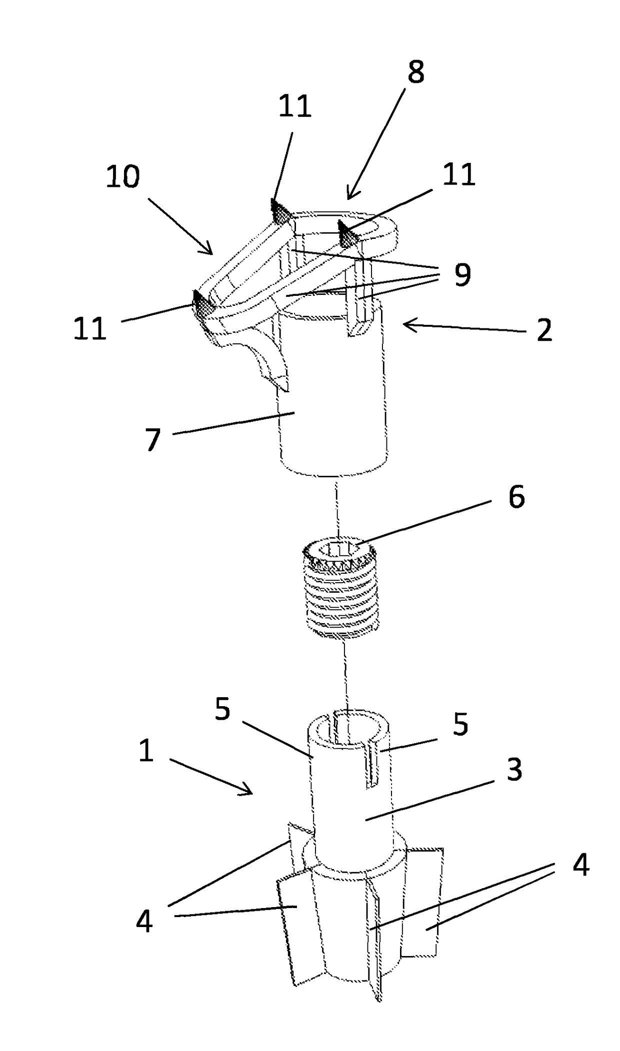
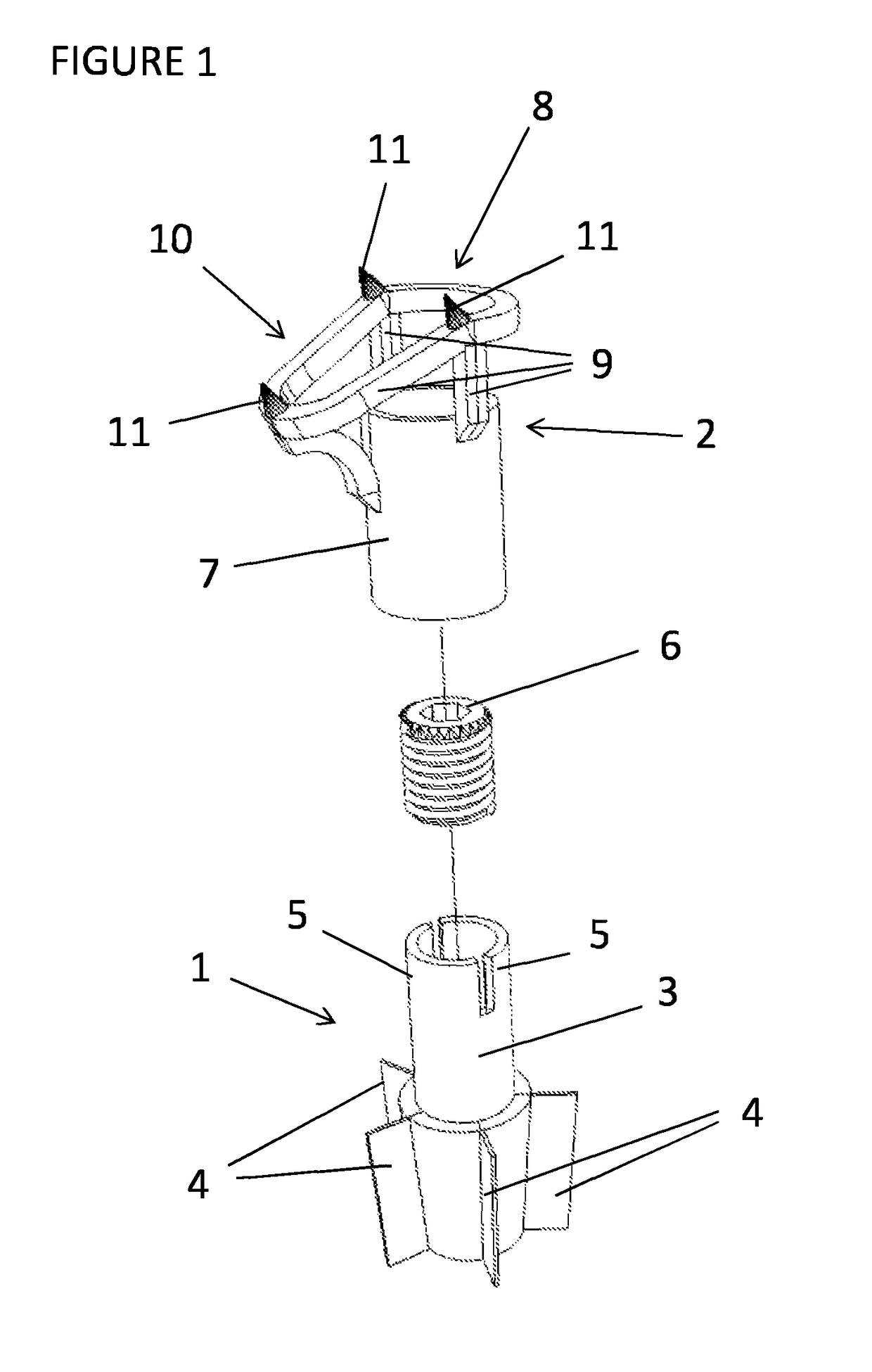
[0077]FIG. 1 shows an embodiment of an implantable medical device according to the invention in disassembled and perspective view. The medical device is configured for treatment of a proximal humerus fracture and comprises a base element 1 to be anchored in the medullar cavity of a humeral shaft of a humerus to be treated and a support element 2 configured to support one or more bone fragments of the humerus.

[0078]The base element 1 comprises a base element body forming a cylindrical extension 3 at its proximal end. The distal end of the base element 1 comprises five anchor fins 4 divided over the circumference of the base element. The anchor fins 4 extend in radial direction and are configured to anchor the base element 1 in the bone of the humeral shaft surrounding the medullar cavity of the humeral shaft.

[0079]The anchor fins 4 are thin walled but stiff elements which provide for proper anchoring of the base element 1 in the medullar cavity. The anchor fins are arranged over a li...


PUM


Abstract

The invention provides an implantable medical device for treatment of a proximal humerus fracture, comprising a base element to be anchored in the medullar cavity of the humeral shaft, a support element to be fixed with respect to the base element, wherein the support element is configured to support one or more bone fragments, wherein the medical device comprises positioning members configured to position the support element with respect to the base element in a range of rotational positions and axial positions, and wherein the medical device comprises a fixation device to fixate the support element, within the range of rotational positions and axial positions, in a desired rotational and axial position with respect to the base element. In an embodiment, the medical device is a non-occluding medical device, wherein after implantation the medullar cavity is not occluded by the medical device due to the open and non canal filling structure of the device preserving blood flow to the reattached bone fragments.

Description

[0001]The present invention relates to a medical device for reconstruction of a humerus for the operative treatment of a proximal humerus fracture.BACKGROUND OF THE INVENTION[0002]Proximal fractures of the humerus are fractures often related to osteoporosis, which occurs frequently in elder people. A proximal fracture of the humerus results in two or more fractured bone fragments. These bone fragments may comprise the humeral head, the humeral shaft, and the greater or lesser tuberosity.[0003]If the bone fragments of the broken humerus are still properly positioned after the fracture, the patient may be successfully treated without surgery. If the bone fragments are dislocated, the position of the bone fragments is often unfavourable so that non-operative treatment would lead to incorrectly or no growing together of the bone fragments.[0004]In those cases the results are generally quite poor and lead to permanent stiffness, pain and limitation of shoulder movements and possibly inva...

Claims


Application Information

Patent Timeline
Patent Type & Authority Patents(United States)
IPC IPC(8): A61F2/38A61B17/68A61B17/72A61B17/74
CPCA61F2/3804A61B17/68A61B17/72A61B17/74A61F2002/3822A61F2220/0008A61B17/7283
Inventor VISSER, CORNELIS PIETER JANBULSTRA, TEAKE HANS
Owner IMPLANT SERVICE BV
Why Eureka
Try Eureka