System and method for visualizing electrophysiology data

a technology of electrophysiology and data, applied in the field of patient electrophysiology visualization, can solve the problems of not being able to receive another stimulus, not being able to fully recover, and unable to conduct a damaged heart muscle, so as to avoid overlapping sequences of electrode acquisition, facilitate viewing ease

Active Publication Date: 2017-05-30
BIOSENSE WEBSTER (ISRAEL) LTD
View PDF26 Cites 56 Cited by
  • Summary
  • Abstract
  • Description
  • Claims
  • Application Information

AI Technical Summary

Benefits of technology

[0049]In some embodiments, the methods include adjusting time scale of the visual representation in response to a user's selected time scale and / or adjusting or limiting the ratio of visual representations to actual cardiac cycles in order to facilitate ease of viewing and avoid overlapping sequences of electrode acquisitions.

Problems solved by technology

During this repolarization phase, the cell is said to be refractory, meaning it has not fully recovered and cannot yet receive another stimulus.
Also, scars from cardiac surgery or damaged heart muscle do not conduct either, and the wave must make its way around the outside edge of these as well.
Thus, the wave of depolarization cannot, in a normal heart, simply continue straight across from the atria to the ventricles.
Should a physician place the catheter in a different location than he or she thinks, or if it should dislodge after being placed in the correct location, the information provided by its electrograms will be inaccurate, leading to delays or even missed or incorrect diagnoses.
The process of building a map—maneuvering the mapping catheter to numerous locations in the heart chamber to sample the electrical data there—takes time.
However, it is still necessary to confirm that the ablation line is truly complete because the flutter may terminate when the tissue is merely damaged and not yet truly ablated.
If remapping is performed for each block assessment with each remapping for a new set of LATs each taking 5-10 minutes to create, these repeated remappings can greatly lengthen the duration of the ablation procedure.
Due to the technical challenges associated with LAT mapping inside the PV, and the fact that the activation sequence changes continuously as more of the tissue is ablated, electrophysiologists almost universally rely on analyzing the Lasso electrograms on the recording system rather than mapping and remapping each change.
However, technical considerations may cause the distal loop to be positioned randomly with its distal and proximal electrodes in any radial orientation.
Due to the technical challenge of positioning the mapping / ablation catheter with sufficient contact to create durable lesions at each location around each pulmonary vein ostium, multiple ablations at the same locations are frequently required, and continual analysis of the Lasso electrograms is critical.
However, activation mapping is rarely done.
Though helpful for creating LAT maps more quickly by taking multiple points at a time, the PentaRay's electrograms are very difficult to recognize by activation pattern alone (see, for example, FIG. 4B).

Method used


Image

  • System and method for visualizing electrophysiology data
  • System and method for visualizing electrophysiology data
  • System and method for visualizing electrophysiology data

Examples


Embodiment Construction

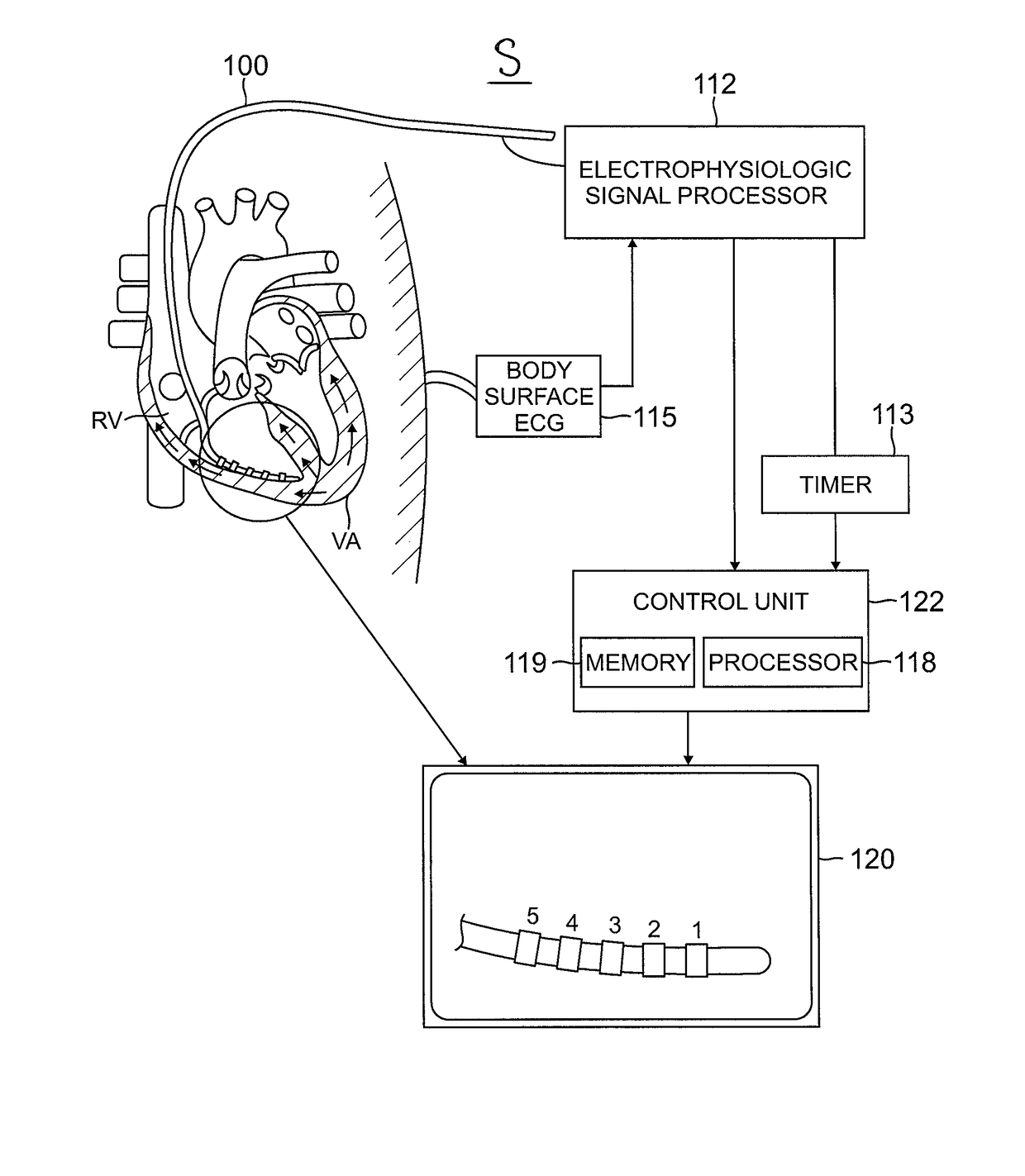
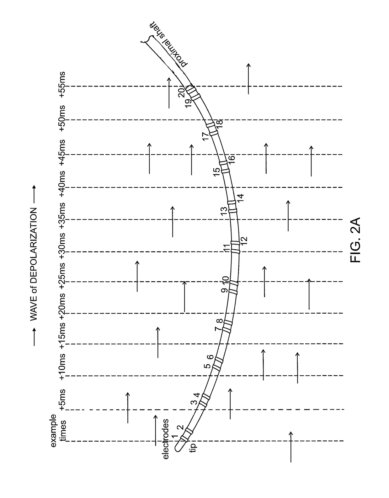
[0095]The present invention is directed to a system and method for real-time visual portrayal of an acquisition sequence of electrodes on a catheter, in particular, real-time visual portrayal of acquisition sequence of electrodes acquiring local activation signals for generating electrograms of a heart. In some embodiments, the visualization of electrode acquisition sequence includes images of the catheter and its electrodes, and visual indicia of electrical propagation along at least an electrical sensing portion of the catheter, including, for example, visual indicia that distinguish acquiring electrode(s) from nonacquiring electrode(s) in real time. In some embodiments, the images include visual indicia of electrical propagation along electrical sensing portions of the catheter, and nonelectrical sensing portion, including, for example, acquiring electrodes and nonconductive tubing extending therebetween. In some embodiments, the visualization of the electrode acquisition sequenc...


PUM


Abstract

A method and system for visualization of electrophysiology information sensed by electrodes on a catheter, includes recording times of electrode signal acquisition, designating a reference electrode signal acquisition, assigning a relative time to each recorded time of electrode signal acquisition relative to the reference electrode signal acquisition, identifying the electrodes with signal acquisition, correlating assigned relative times to identified electrodes to generate a sequence of electrode signal acquisitions, and generating a visual representation of the sequence of electrode signal acquisitions generating a visual representation with a graphical image of the electrodes, wherein individual electrodes are visually marked to represent the sequence of electrode signal acquisitions.

Description

FIELD OF INVENTION[0001]This invention relates generally to visualization of electrical activity of a patient, and more particularly to spatial visualization of electrophysiology data for a patient as detected by a catheter.BACKGROUND[0002]Cardiac arrhythmias are the leading cause of death in the United States. Normally, electrical acquisition signals are conducted in an orderly way through the atrium and into the ventricle, passing each point in the heart only once in each heart cycle. Electrical acquisition signals at different locations in the heart are well correlated, taking into account normal propagation delays from one region of the heart to another. In response to local activation signals, the atrial muscle fibers contract in proper synchrony to pump blood through the atrium.[0003]The electrical conduction system of the heart provides the rhythm, or sequence of contraction of the heart muscle, so it may most efficiently pump blood to the rest of the body. Electrophysiologis...

Claims


Application Information

Patent Timeline
Patent Type & Authority Patents(United States)
IPC IPC(8): A61B5/044A61B5/042A61B5/04A61B5/00A61B5/0432A61B5/296
CPCA61B5/044A61B5/0422A61B5/04012A61B5/0432A61B5/7246A61B5/318A61B5/316A61B5/287A61B5/333A61B5/339
Inventor SEVERINO, THOMAS MICHAEL
Owner BIOSENSE WEBSTER (ISRAEL) LTD
Why Eureka
Try Eureka