Method and apparatus for tissue disease diagnosis

a tissue disease and optical diagnosis technology, applied in the field of tissue abnormality diagnosis, can solve the problems of slow growth of tumors, inability to achieve simultaneous high labor intensity of analyzing pap smears, and achieve the effect of high sensitivity and specificity, high labor intensity and high specificity

Active Publication Date: 2018-02-27
BIOP MEDICAL
View PDF16 Cites 0 Cited by
  • Summary
  • Abstract
  • Description
  • Claims
  • Application Information

AI Technical Summary

Benefits of technology

[0024]Additional features and advantages of the invention will become apparent from the following drawings and description.

Problems solved by technology

Second, these are slow growing tumors.
Unfortunately, the Pap smear is unable to achieve a concurrently high sensitivity and high specificity due to both sampling and analysis errors.
Additionally, analyzing Pap smears is extremely labor intensive and requires highly trained professionals.
Extensive training is necessary in order for a practitioner to perform colposcopy and its diagnosis accuracy is variable and limited, even in expert hands.
Disadvantageously, EIS has positive predictive value (PPV) of only 72% and is an expensive process.

Method used


Image

  • Method and apparatus for tissue disease diagnosis
  • Method and apparatus for tissue disease diagnosis
  • Method and apparatus for tissue disease diagnosis

Examples


Embodiment Construction

[0050]Before explaining at least one embodiment in detail, it is to be understood that the invention is not limited in its application to the details of construction and the arrangement of the components set forth in the following description or illustrated in the drawings. The invention is applicable to other embodiments being practiced or carried out in various ways. Also, it is to be understood that the phraseology and terminology employed herein is for the purpose of description and should not be regarded as limiting.

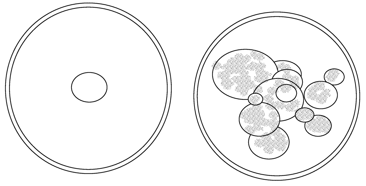
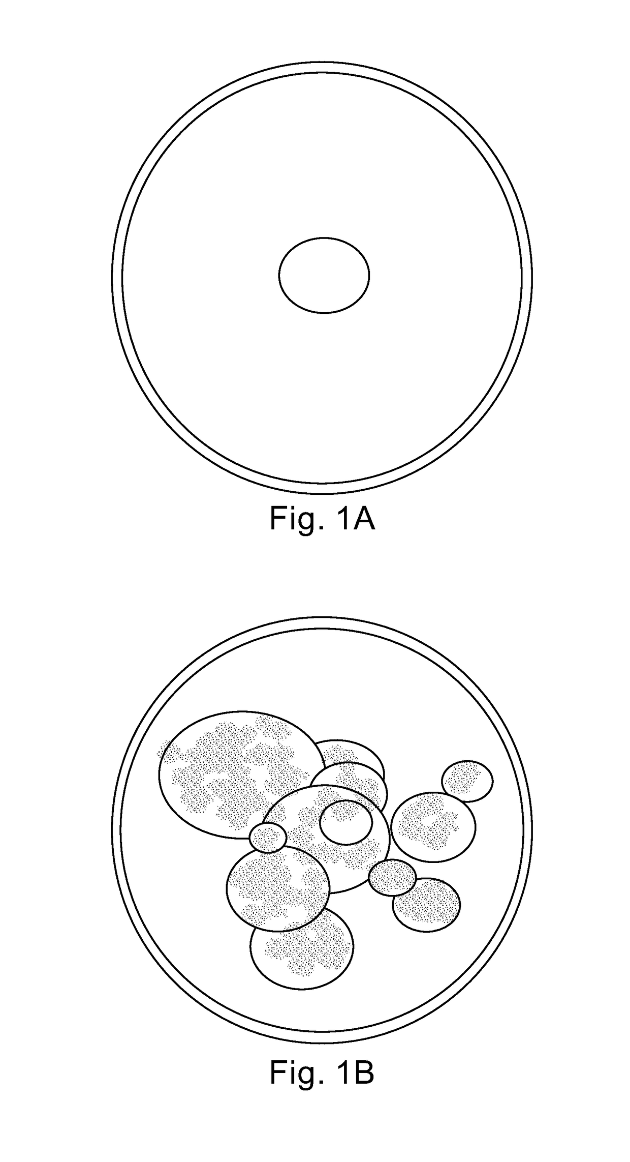
[0051]A main difference between cervical cancer cells and normal cervical cells is their nuclear structure. Normal cells have a single nucleus, as shown in FIG. 1A, with a narrow range of sizes. The nucleus is generally round, oval or bean shaped. Pre-cancer and cancer cells have multiple nuclei, as shown in FIG. 1B, with a wide range of shapes and sizes. As a result, cancer cells scatter light by larger angles than healthy cells. Another reason for scattering of th...


PUM


Abstract

A tissue diagnosis apparatus constituted of: a broad band light source arranged to irradiate a target area with broad band light; a narrow band light source arranged to irradiate the target area with narrow band light; at least one light sensor arranged to: sense the broad and narrow band light after interaction with the target area; a color sensor arranged to sense the broad band light after interaction with the target area; and a housing, the broad band light source, narrow band light source, light sensor and color sensor situated within the housing, wherein a control circuitry is arranged to: determine the image abnormality of the sensed broad band light, the spatial scattering of the sensed narrow band light and the chromatic impact of the interaction of the broad band light with the target area; and output a signal responsive to the identified image abnormality, spatial scattering and chromatic impact.

Description

CROSS-REFERENCE TO RELATED APPLICATIONS[0001]This application claims priority from U.S. Provisional Patent Application Ser. No. 61 / 596,262, filed Feb. 8, 2012 and entitled “DIAGNOSTIC SYSTEM FOR DETECTION OF ABNORMAL CONDITION OF CERVICAL TISSUE”, the entire contents of each of which are incorporated herein by reference.FIELD OF THE INVENTION[0002]The invention relates generally to the field of tissue abnormality diagnosis and in particular to a method and apparatus for optical diagnosis of tissue diseases.BACKGROUND OF THE INVENTION[0003]Cervical cancer is one of the common neoplasms of the female genital tract. Cervical cancer is the second malignancy in women worldwide and is one of the leading causes of women death in the third world. Early diagnosis of abnormal cells in the cervix prevents deterioration into fully cervical cancer and thus reduces morbidity and mortality. The pre-cancerous state is called Squamous Intraepithelial Lesion (SIL), and has two grades: low grade SIL a...

Claims


Application Information

Patent Timeline
Patent Type & Authority Patents(United States)
IPC IPC(8): A61B5/00A61B1/00A61B1/303A61B1/06
CPCA61B5/4331A61B1/00009A61B5/0084A61B1/303A61B5/0062A61B1/0638
Inventor LANDESMAN, ILAN
Owner BIOP MEDICAL
Why Eureka
Try Eureka