Application of hepatocyte model derived in vitro by stem cell
A technology of hepatocytes and stem cells, applied in the fields of developmental biology and pharmacology, can solve problems such as the application of stem cell-derived hepatocyte models, etc.
- Summary
- Abstract
- Description
- Claims
- Application Information
AI Technical Summary
Problems solved by technology
Method used
Image

Examples
Embodiment 1
[0017] Example 1: The ES cell in vitro derivation liver cell model is constructed by the following steps
[0018] 1. Hanging drop culture
[0019] ES cells were digested with trypsin to make a cell density of 2×10 4 cells / ml of single-cell suspension, inoculated on the inner surface of the lid of a 10-cm diameter petri dish with 30 μl, added 10 ml of D-Hanks solution into the petri dish, and then carefully covered the lid with many small drops to make the lid The small drops on the surface were hung upside down to form hanging drops (each hanging drop contained about 600 cells), and cultured in an incubator for 5 days. The differentiation medium was high-glucose DMEM, containing 0.1 mmol·L -1 β-mercaptoethanol, non-essential amino acids and 20% fetal bovine serum without LIF factor.
[0020] 2. Adhesive culture
[0021] Transfer the embryoid body (embryoid body, EB) formed by hanging drop culture with a pipette under the microscope to a 24-well plate coated with gelatin, a...
Embodiment 2
[0024] Example 2: In vitro derivation of ES cells into liver cells Mitochondrial membrane potential changes
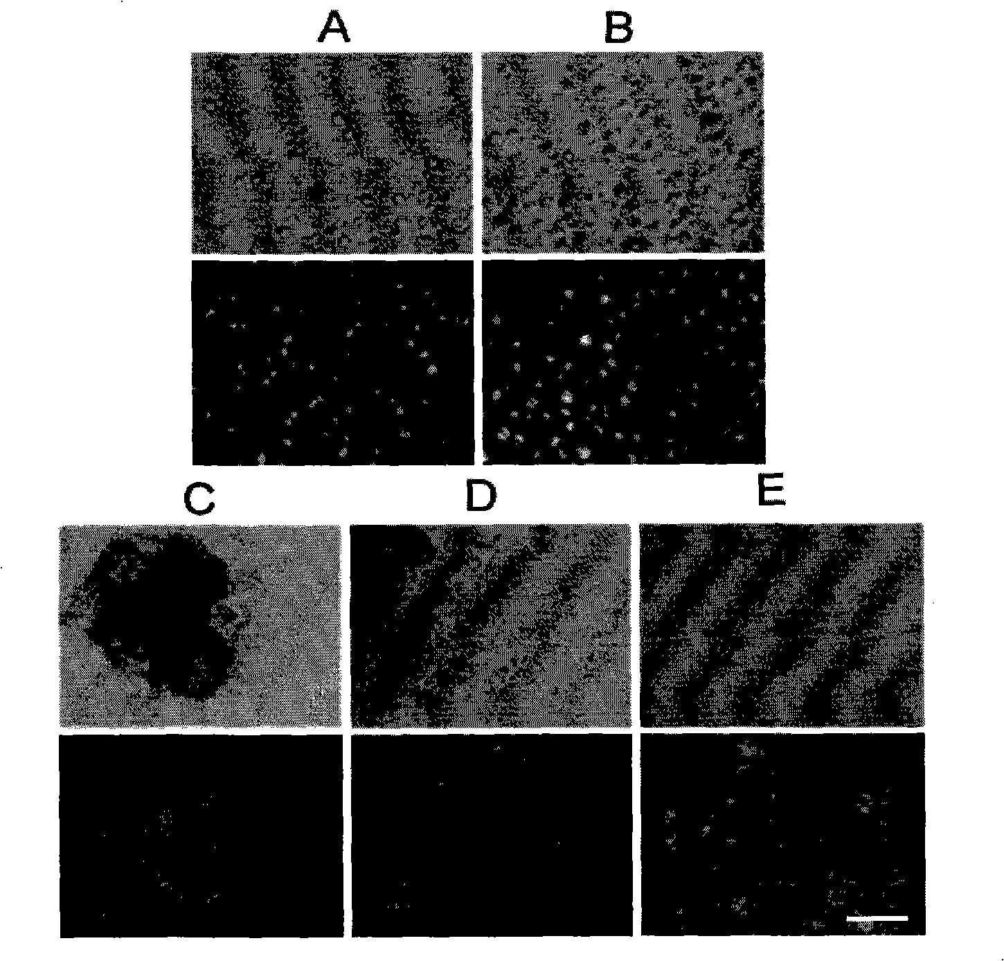
[0025] ES cells were collected, EBs were digested into single cells, EBs were adhered to the wall and differentiated into hepatocyte samples, added JC-1 dye and incubated for 15 minutes, washed three times with PBS, and observed under a fluorescent inverted microscope.
[0026] The results showed that almost all ES cells exhibited green fluorescence, suggesting that the mitochondrial membrane potential was low; EB digested cells showed red fluorescence, while almost all hepatocytes cultured by adherent differentiation showed red fluorescence, which suggested that with the Differentiation and maturation, the cell membrane potential gradually increased with the maturation of liver cell mitochondria. see figure 2 , where (A) a single ES cell, ×100; (B) a single cell after digested EB, ×100; (C) the 12th day of EB adherent culture differentiation, ×50; (D) the 12th day o...
Embodiment 3
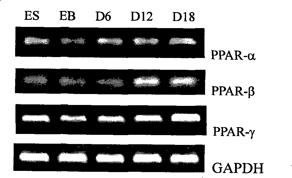
[0027] Example 3: Expression of PPARs family genes when ES cells are derivatized into hepatocytes in vitro
[0028] Collect ES cells, EBs, and hepatocytes on days 6, 12, and 18 of adherent differentiation, and extract total mRNA according to the instructions of Trizol reagent. See Table 1 for primer sequences and experimental conditions:
[0029] Table 1. Primer sequences and PCR reaction conditions
[0030]
[0031] PCR products were electrophoresed on 1.5% agarose gel, observed and photographed with a gel imaging system.
[0032] see image 3 , the experimental results showed that PPAR-α, PPAR-β and PPAR-γ genes were expressed during the differentiation of ES cells into liver cells, and all of them had a certain degree of up-regulation trend.
PUM
 Abstract
Description
Claims
Application Information
 - R&D Engineer
- R&D Manager
- IP Professional
- Industry Leading Data Capabilities
- Powerful AI technology
- Patent DNA Extraction
Browse by: Latest US Patents, China's latest patents, Technical Efficacy Thesaurus, Application Domain, Technology Topic.
© 2024 PatSnap. All rights reserved.Legal|Privacy policy|Modern Slavery Act Transparency Statement|Sitemap